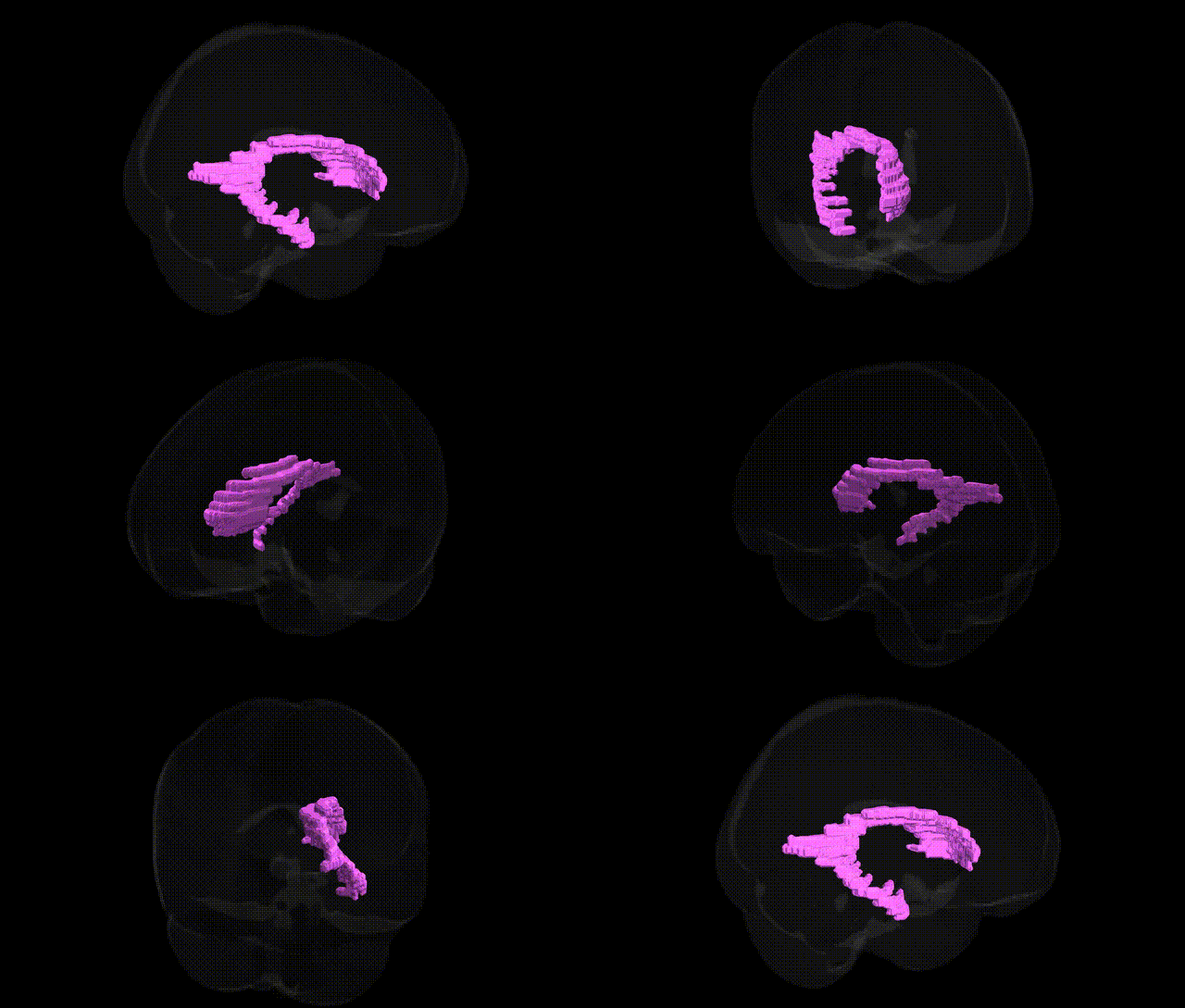
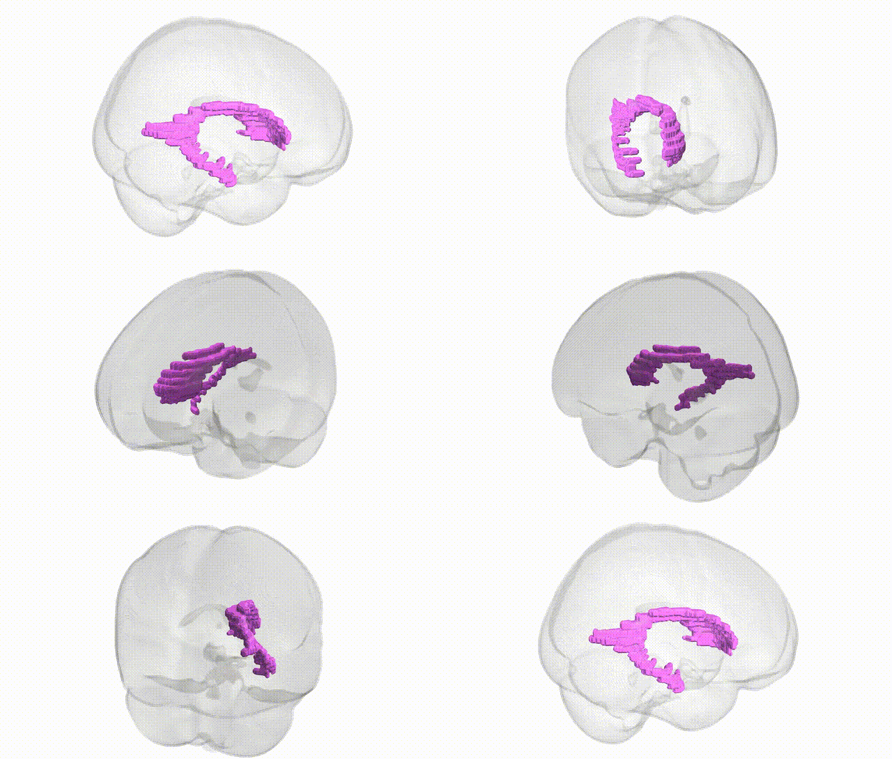
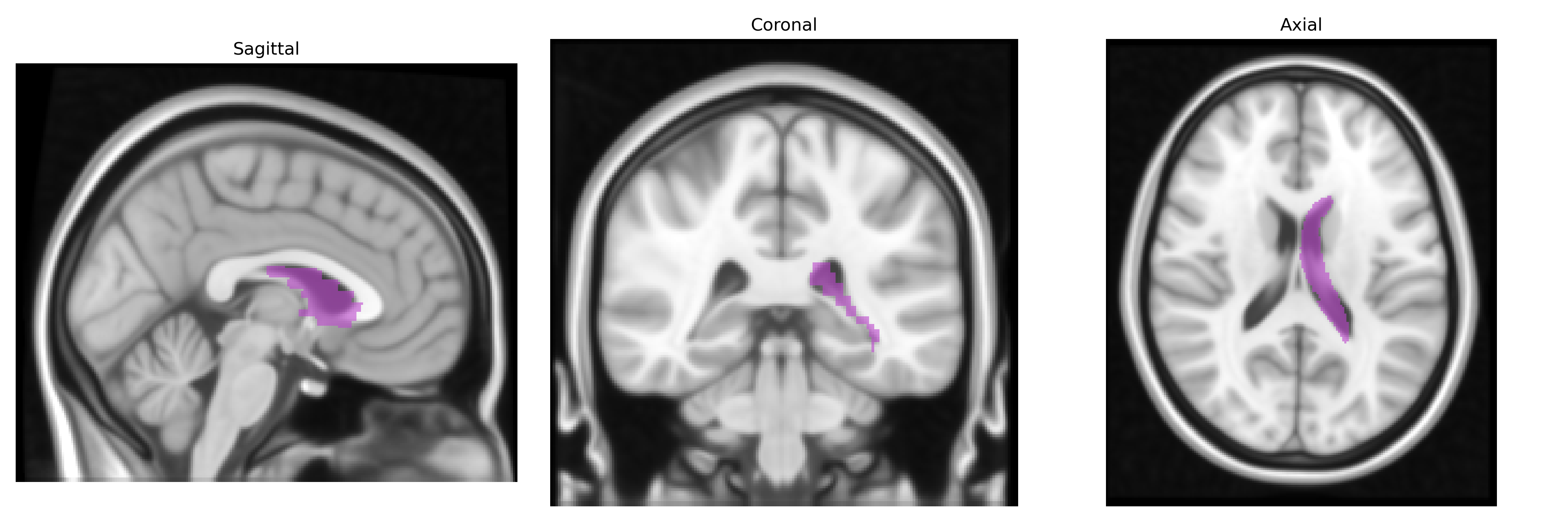
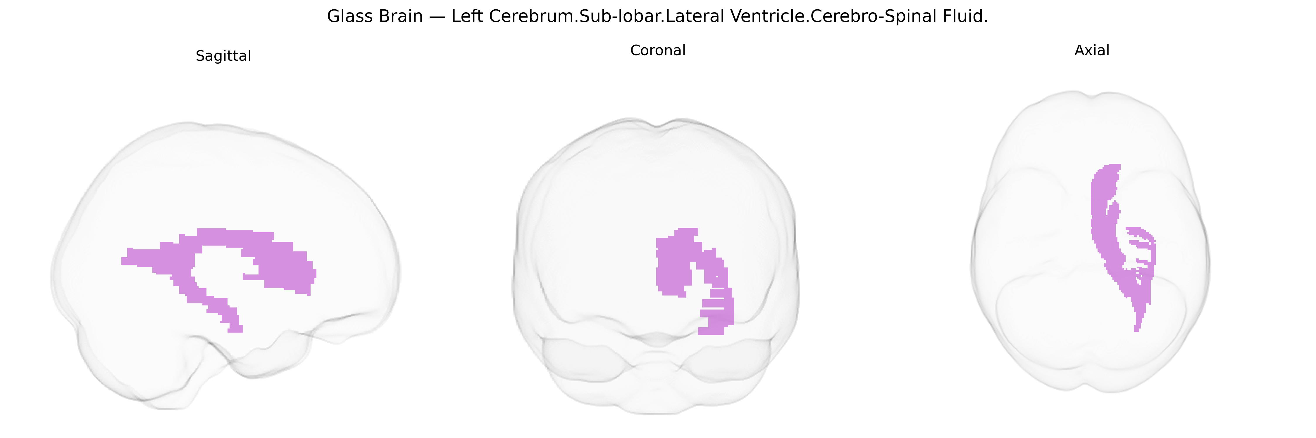
The bilateral Left Cerebrum.Sub-lobar.Lateral Ventricle.Cerebro-Spinal Fluid region, as defined in the Talairach 1 mm Atlas, corresponds to the cerebrospinal fluid (CSF)–filled cavities of the lateral ventricles within the left cerebral hemisphere’s subcortical (sub-lobar) territory. These ventricles are part of the ventricular system that produces, contains, and circulates CSF, which cushions the brain, maintains intracranial pressure, and participates in metabolic waste clearance. Anatomically, the lateral ventricles extend through the frontal, parietal, temporal, and occipital lobes, and their CSF content serves as an important landmark in neuroimaging for delineating adjacent deep gray and white matter structures, including the caudate nucleus, thalamus, and corpus callosum. There is no direct Wikipedia article for this exact label; for a closely related structure, see Lateral ventricle.
The bilateral left sub-lobar lateral ventricles, which contain cerebrospinal fluid rather than neuronal tissue, are primarily implicated in genetic studies through genome-wide association analyses of ventricular volume and related brain structural phenotypes. Numerous GWAS have shown that lateral ventricular enlargement is highly heritable and associated with variants in genes involved in neurodevelopment, extracellular matrix organization, and neurodegeneration, including loci near/in APOE (particularly ε4), MAPT, and other Alzheimer’s disease–related genes, as well as genes linked to schizophrenia, bipolar disorder, and major depression. Ventricular volume–associated variants have also been reported near genes influencing intracranial volume (e.g., loci at 17q21.31 and 12q24), reflecting generalized brain atrophy and neurodevelopmental processes. Enlargement of the lateral ventricles has been repeatedly associated, at the genetic level, with neurodegenerative diseases (especially Alzheimer’s disease), psychotic disorders, and overall brain aging, with polygenic risk scores for these conditions correlating with increased ventricular volume. Although the Talairach 1 mm atlas designates this region anatomically, genetic associations are typically reported for global or lateral ventricular volume rather than the specific “left sub-lobar lateral ventricle CSF” label, and current findings thus reflect ventricular size as a biomarker of genetically influenced brain atrophy and neurodevelopmental variation rather than a trait of the CSF compartment itself.
Overview generated by GPT-4o (2026).
Region ID: 173
Hemisphere: bilateral
Atlas: Talairach labels 1mm
Full Quality Version: Download MP4
Full Quality Version: Download MP4
Wali Sidiqyar*, Gaurav Rudravaram*, Elyssa M. McMaster, Trent M. Schwartz, Adam M. Saunders, Kurt G. Schilling, Bennett A. Landman "Introducing SPINS: A Shared Public Visualization Library of Neuroanatomical Structures." Medical Imaging with Deep Learning- short paper
This resource is licensed under CC0 1.0 Universal (Public Domain).