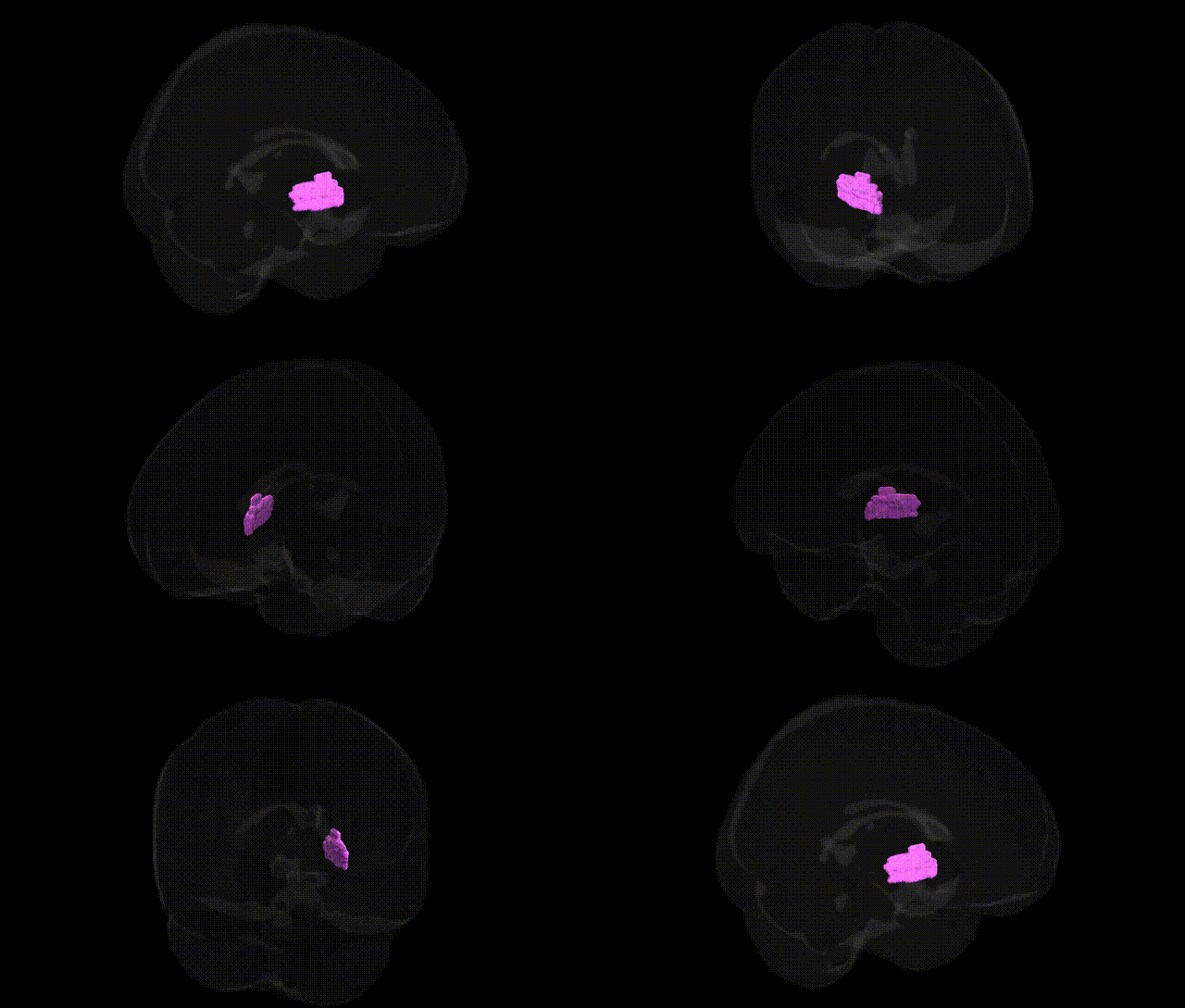
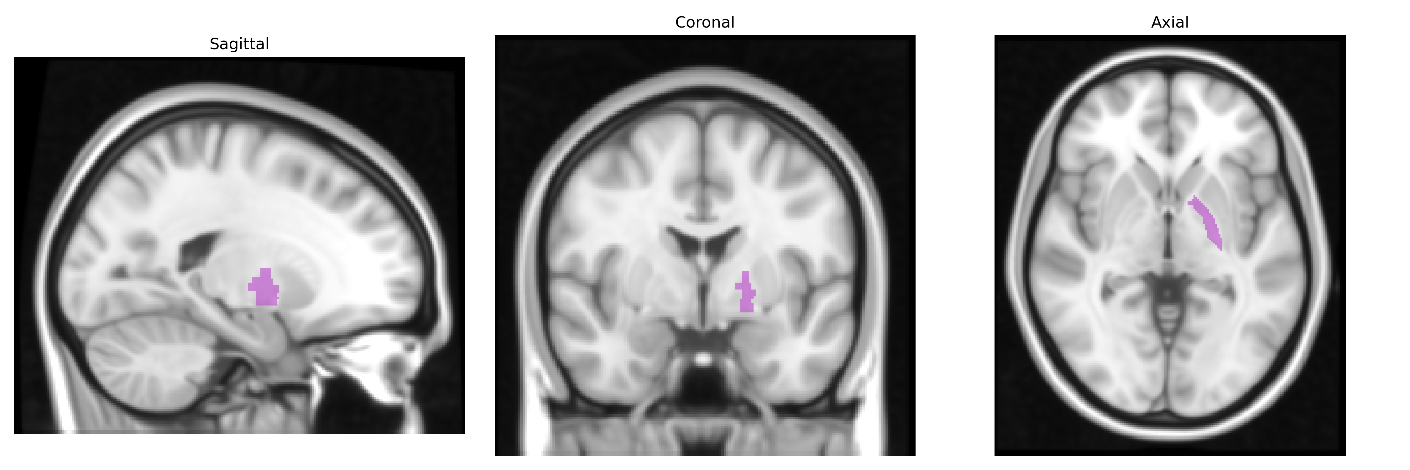
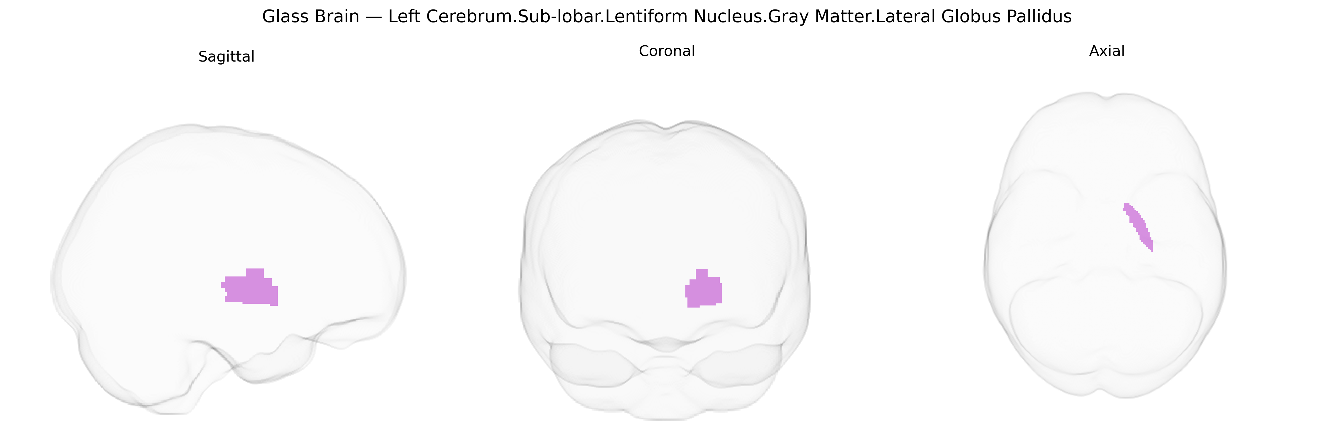
The bilateral Left Cerebrum.Sub-lobar.Lentiform Nucleus.Gray Matter.Lateral Globus Pallidus corresponds to the lateral segment of the globus pallidus, a major inhibitory nucleus of the basal ganglia located deep within the subcortical white matter of the cerebral hemispheres. This structure forms part of the lentiform nucleus alongside the putamen and participates critically in motor control, particularly in the indirect pathway, where it receives input from the striatum and projects to the subthalamic nucleus. Neurons in the lateral globus pallidus are predominantly GABAergic, exerting tonic inhibitory influence that modulates thalamocortical activity and helps regulate movement initiation, amplitude, and suppression of unwanted motor patterns. Dysfunction or degeneration of this region is implicated in movement disorders such as Parkinson’s disease and Huntington’s disease, reflecting its central role in basal ganglia circuitry. Globus pallidus
The lateral globus pallidus, a core node of the basal ganglia within the lentiform nucleus, has been implicated in multiple genetic and GWAS-based associations through its roles in motor control, reward processing, and cognitive-emotional regulation. Variants in genes affecting dopaminergic and basal ganglia circuitry—such as DRD2, DRD3, COMT, and several glutamatergic and GABAergic pathway genes—have been linked to functional and structural changes in this region, often via imaging genetics studies that report altered pallidal volume, connectivity, or activation. Large-scale GWAS of subcortical brain volumes (e.g., ENIGMA, UK Biobank) have identified common variants near or within genes involved in neurodevelopment, axon guidance, and synaptic plasticity (including loci in or near DCC, CELSR3, and other neurodevelopmental genes) that associate with globus pallidus volume and morphology. Clinically, genetic risk variants for movement disorders such as Parkinson’s disease, dystonia, and Huntington’s disease (e.g., SNCA, LRRK2, GBA, HTT) converge on basal ganglia circuits that prominently involve the lateral globus pallidus, while polygenic risk for psychiatric disorders—including schizophrenia, major depression, and obsessive-compulsive disorder—has been linked to structural and functional alterations in pallidal regions through imaging-genetics approaches. Additionally, alcohol and substance use GWAS have implicated dopaminergic and striato-pallidal pathway genes, consistent with the lateral globus pallidus’ role in reward and habit circuits, though associations are often at the circuit or network level rather than uniquely localized to this sub-lobar structure.
Overview generated by GPT-4o (2026).
Region ID: 452
Hemisphere: bilateral
Atlas: Talairach labels 1mm
Full Quality Version: Download MP4
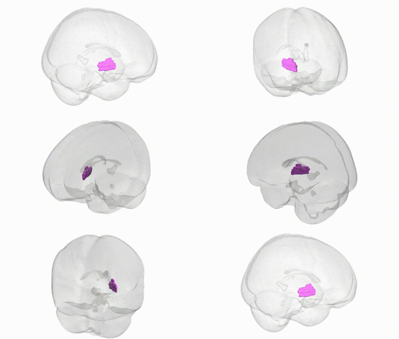
Full Quality Version: Download MP4
Wali Sidiqyar*, Gaurav Rudravaram*, Elyssa M. McMaster, Trent M. Schwartz, Adam M. Saunders, Kurt G. Schilling, Bennett A. Landman "Introducing SPINS: A Shared Public Visualization Library of Neuroanatomical Structures." Medical Imaging with Deep Learning- short paper
This resource is licensed under CC0 1.0 Universal (Public Domain).