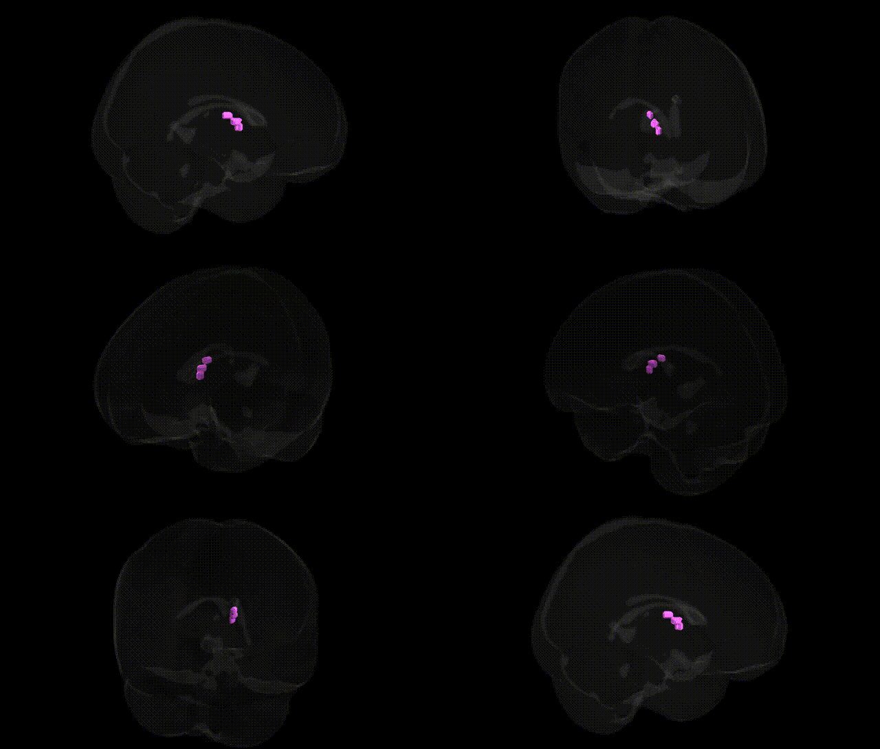
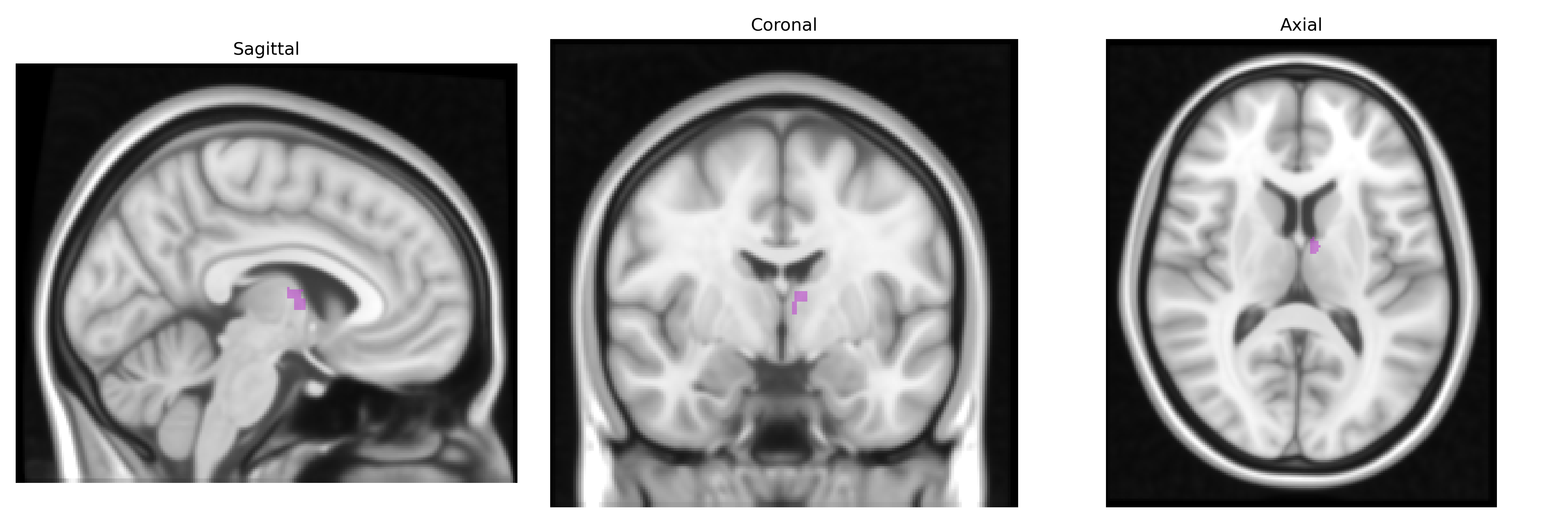
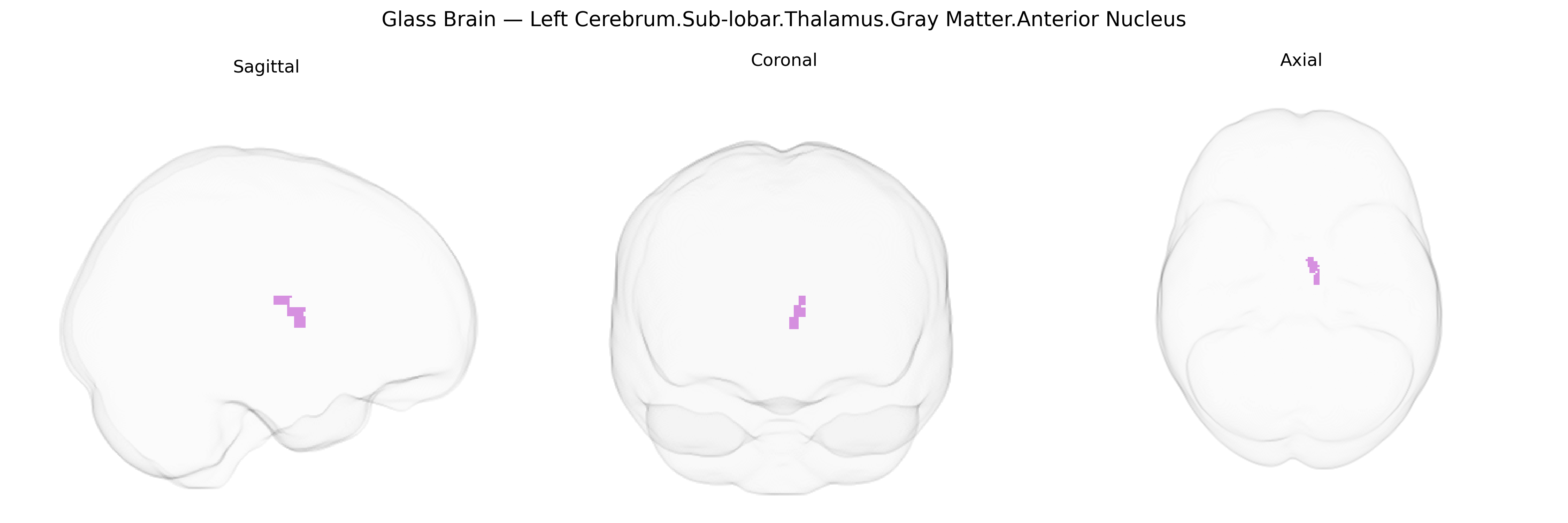
The bilateral Left Cerebrum.Sub-lobar.Thalamus.Gray Matter.Anterior Nucleus corresponds to the anterior nuclei of the thalamus, a key relay complex within the limbic system located in the medial and rostral portion of the dorsal thalamus. These nuclei receive major afferent input from the mammillary bodies via the mammillothalamic tract and from the hippocampal formation through the fornix, and project primarily to the cingulate gyrus and other limbic cortical areas. Functionally, the anterior thalamic nuclei are implicated in episodic memory, spatial navigation, emotional processing, and aspects of attention, often working in concert with the hippocampus and cingulate cortex as part of the Papez circuit. Lesions in this region are associated with memory impairments and diencephalic amnesia, highlighting its importance in declarative memory networks. Anterior nuclei of thalamus
The bilateral anterior nucleus of the thalamus, within the sublobar gray matter of the left (and right) cerebrum as defined in the Talairach 1 mm atlas, has been indirectly implicated in multiple genetic and GWAS findings through its roles in limbic circuitry, memory, and cortico-thalamo-cortical networks rather than by many region-specific association studies. Polygenic and GWAS analyses of thalamic volume and shape (e.g., ENIGMA and UK Biobank–based studies) have identified common variants in genes such as MAPT, NPTX1, and loci near BCL11B, TCF4, and GRIN2A that influence overall thalamic size, specific thalamic nuclei, or thalamocortical connectivity, with downstream effects most notable in anterior and mediodorsal nuclei linked to cognition and psychiatric risk. Thalamic GWAS also overlap with schizophrenia, bipolar disorder, and major depressive disorder polygenic risk, highlighting shared variants in synaptic, glutamatergic, and neurodevelopmental genes that track with structural differences in anterior and mediodorsal thalamic regions. In addition, genetic studies of temporal lobe epilepsy and limbic epilepsies, including those examining responsiveness to deep brain stimulation of the anterior nucleus, suggest that variants in genes governing excitability (e.g., SCN and GABA receptor genes) and synaptic plasticity contribute to individual differences in anterior thalamic structure and seizure networks, though effects are typically network-wide rather than nucleus-specific. Finally, imaging-genetics work links APOE and other Alzheimer’s disease risk loci to anterior thalamic and connected limbic structural changes in late-life, consistent with the nucleus’s involvement in memory circuits, but the field still lacks large, finely parcellated GWAS focused exclusively on the bilateral anterior thalamic nucleus as delineated in the Talairach 1 mm atlas.
Overview generated by GPT-4o (2026).
Region ID: 686
Hemisphere: bilateral
Atlas: Talairach labels 1mm
Full Quality Version: Download MP4
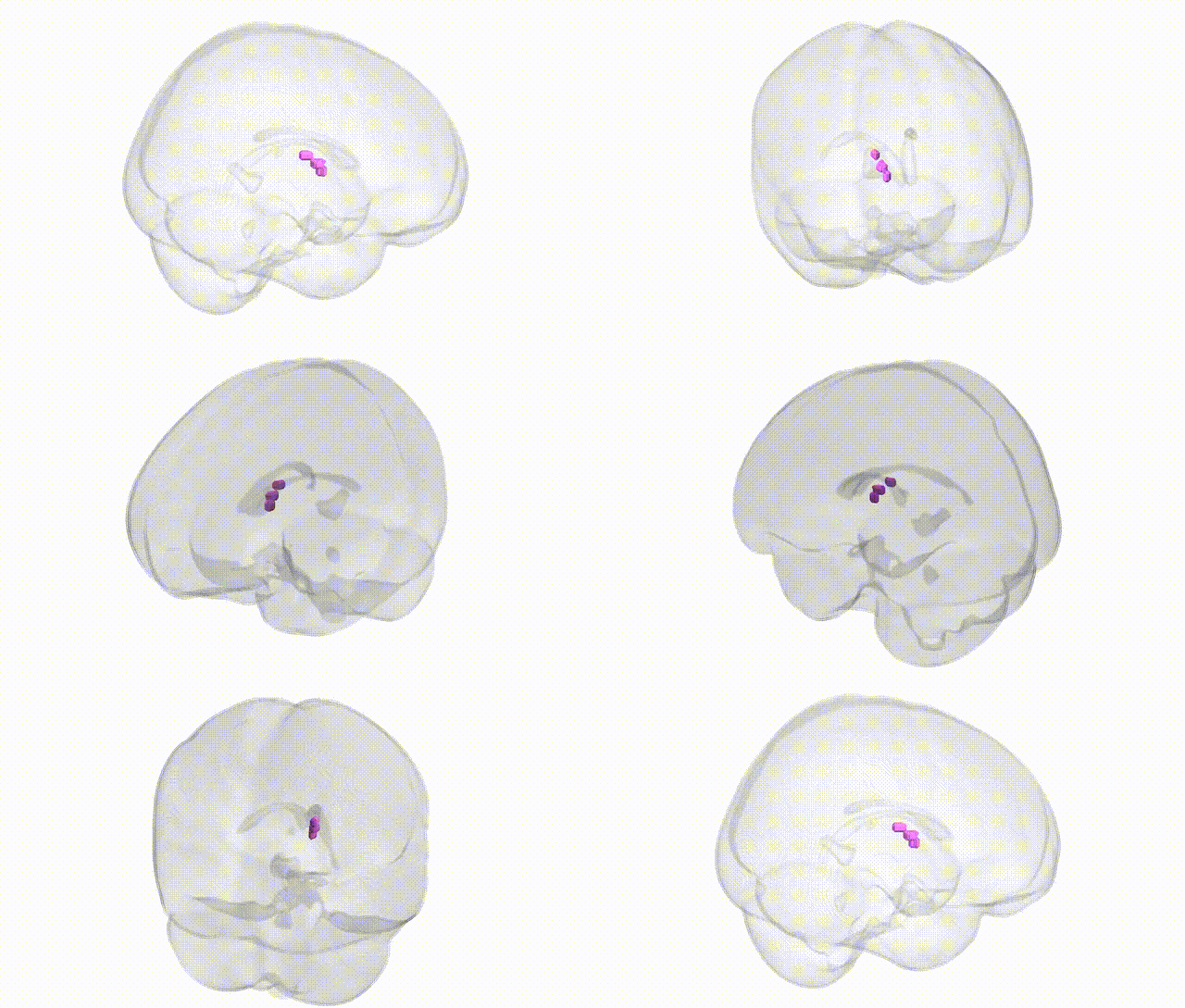
Full Quality Version: Download MP4
Wali Sidiqyar*, Gaurav Rudravaram*, Elyssa M. McMaster, Trent M. Schwartz, Adam M. Saunders, Kurt G. Schilling, Bennett A. Landman "Introducing SPINS: A Shared Public Visualization Library of Neuroanatomical Structures." Medical Imaging with Deep Learning- short paper
This resource is licensed under CC0 1.0 Universal (Public Domain).