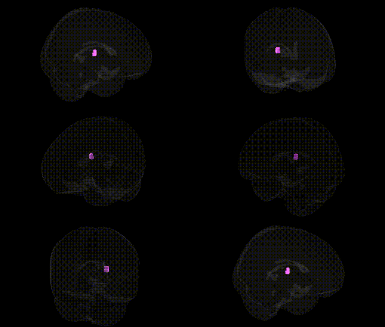
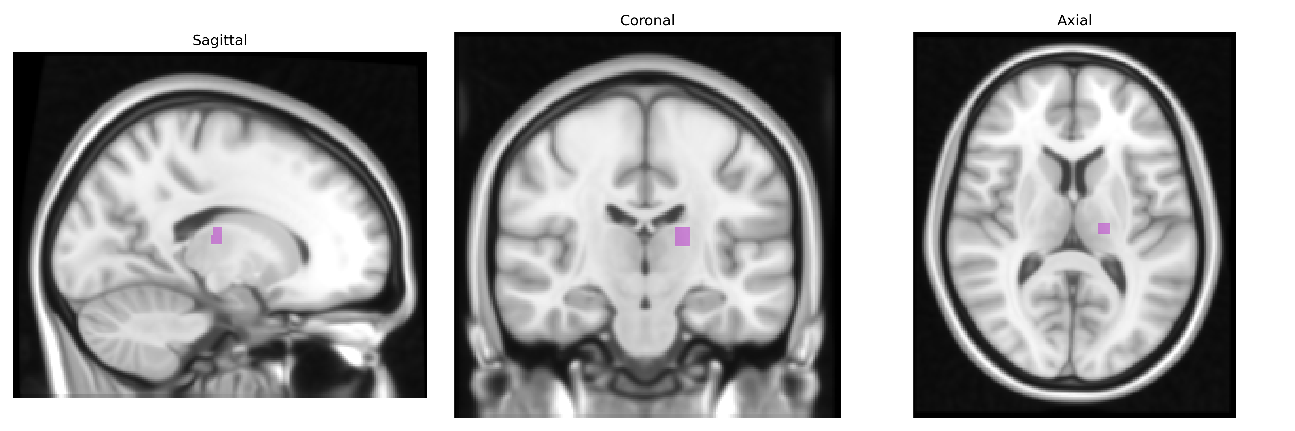
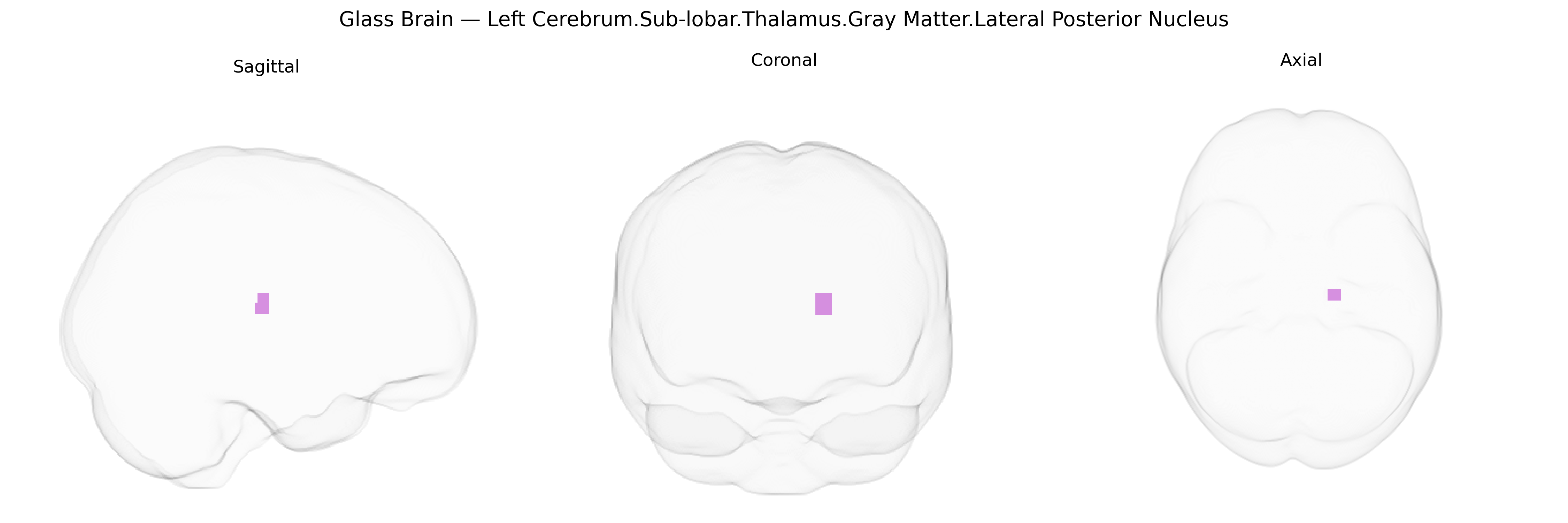
The bilateral Left Cerebrum.Sub-lobar.Thalamus.Gray Matter.Lateral Posterior Nucleus corresponds to the lateral posterior (LP) nucleus of the thalamus in both hemispheres, a higher-order associative thalamic relay located in the subcortical gray matter of the dorsal thalamus. This nucleus is closely related to and sometimes grouped with the pulvinar complex, sharing extensive reciprocal connections with parietal, occipital, and temporal association cortices. Functionally, the LP nucleus participates in visuospatial attention, integration of multimodal sensory information, and modulation of cortical processing important for perception and higher-order cognition. Through its widespread corticothalamocortical loops, it contributes to coordination of sensory-driven behavior and attentional orienting, and is implicated in distributed networks supporting visual processing and sensorimotor integration. There is no direct link for this specific nucleus; see Thalamus.
The lateral posterior nucleus of the thalamus, a higher-order associative relay connected with parietal and temporal cortices, has been implicated in several genetic and neuroimaging-genetics findings, although few GWAS target this subnucleus specifically. Large-scale structural and functional MRI GWAS (e.g., ENIGMA, UK Biobank) have identified common variants near genes involved in neurodevelopment (such as NPTN, DPYSL5, and various synaptic and axon-guidance genes) associated with thalamic volume and thalamo-cortical connectivity, which would encompass or influence the lateral posterior nucleus region. More broadly, polygenic architectures for schizophrenia, bipolar disorder, and major depressive disorder show enrichment in thalamic networks, and case–control imaging-genetics studies report thalamic volume or connectivity differences linked to risk alleles in genes including CACNA1C, ZNF804A, and MIR137-related loci. Thalamic associative nuclei, including lateral posterior, have also been implicated through GWAS of cognitive traits (general intelligence, processing speed) and educational attainment, where thalamic volume and integrity partially mediate the effects of polygenic scores. In movement and neurodegenerative disorders, variants in genes such as LRRK2 and MAPT have been associated with thalamic atrophy or altered thalamic connectivity patterns, while in epilepsy, risk variants affecting thalamocortical circuitry (e.g., in GABRA2 and other GABAergic or glutamatergic genes) are linked to network-level changes that involve the lateral posterior and neighboring nuclei. Overall, genetic associations for this specific Talairach-defined lateral posterior thalamic region are inferred primarily from broader thalamic or thalamo-parietal imaging-genetics and disorder-risk studies rather than nucleus-specific GWAS.
Overview generated by GPT-4o (2026).
Region ID: 725
Hemisphere: bilateral
Atlas: Talairach labels 1mm
Full Quality Version: Download MP4
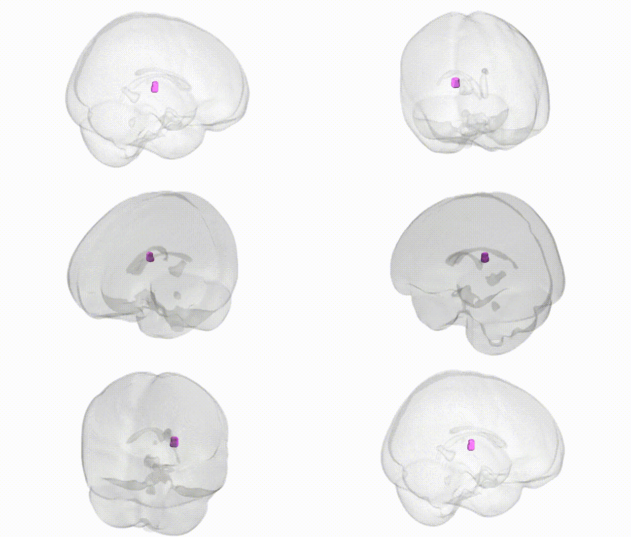
Full Quality Version: Download MP4
Wali Sidiqyar*, Gaurav Rudravaram*, Elyssa M. McMaster, Trent M. Schwartz, Adam M. Saunders, Kurt G. Schilling, Bennett A. Landman "Introducing SPINS: A Shared Public Visualization Library of Neuroanatomical Structures." Medical Imaging with Deep Learning- short paper
This resource is licensed under CC0 1.0 Universal (Public Domain).