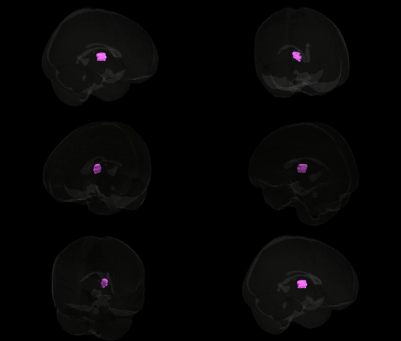
The bilateral Left Cerebrum.Sub-lobar.Thalamus.Gray Matter.Medial Dorsal Nucleus corresponds to the mediodorsal (medial dorsal) nucleus of the thalamus, a higher-order relay and association nucleus situated in the medial thalamus and interconnected extensively with prefrontal, orbitofrontal, and limbic cortical regions. It plays a key role in executive functions, working memory, decision-making, and emotional processing by integrating limbic inputs (including from the amygdala and basal forebrain) with prefrontal output, thereby contributing to goal-directed behavior and affective regulation. Lesions or dysfunction in this nucleus have been associated with cognitive impairments, especially in attention and memory, as well as changes in personality and mood seen in various neuropsychiatric and neurodegenerative disorders. There is no direct link for this exact nuclear subdivision labeling, but a related structure is the Medial dorsal nucleus of thalamus.
The medial dorsal nucleus of the thalamus, a key sub-lobar gray matter region within the left cerebrum, has been repeatedly implicated in genetic studies of cognitive, psychiatric, and neurodegenerative phenotypes. GWAS of brain structure have identified common variants near genes involved in neurodevelopment, synaptic signaling, and axon guidance (e.g., genes in glutamatergic and GABAergic pathways, cell-adhesion molecules, and transcription factors) that influence thalamic volume and microstructure, with bilateral and medial dorsal subregional effects observed in some imaging-genetics consortia such as ENIGMA and UK Biobank–based analyses. Medial dorsal thalamic morphology and connectivity show heritability and have been associated with risk variants for schizophrenia (including loci in the MHC region and other synaptic genes), bipolar disorder, and major depressive disorder, consistent with the nucleus’s role in fronto-thalamic circuits underlying cognition and affect; polygenic risk scores for these disorders often correlate with altered thalamic volume or functional coupling. In autism spectrum disorder and ADHD, GWAS and candidate gene studies have reported thalamic involvement in circuits affected by risk variants (e.g., in genes regulating synapse formation and neurotransmission), with some imaging-genetic work linking higher polygenic risk to developmental changes in thalamic nuclei, including the medial dorsal nucleus. Additionally, variants in genes related to neurodegenerative disease (such as APOE and other Alzheimer’s disease loci) and to sleep/circadian traits show associations with thalamic structure and activity, reflecting the medial dorsal nucleus’s role in memory and arousal networks, though findings are often regionally distributed across thalamic subnuclei rather than exclusive to the medial dorsal nucleus. Overall, genetic influences on this region are typically detected via imaging-genetics rather than single-region GWAS, and most associations emerge in the context of broader thalamic and fronto-striato-limbic circuitry implicated in psychiatric and cognitive traits.
Overview generated by GPT-4o (2026).
Region ID: 636
Hemisphere: bilateral
Atlas: Talairach labels 1mm
Full Quality Version: Download MP4
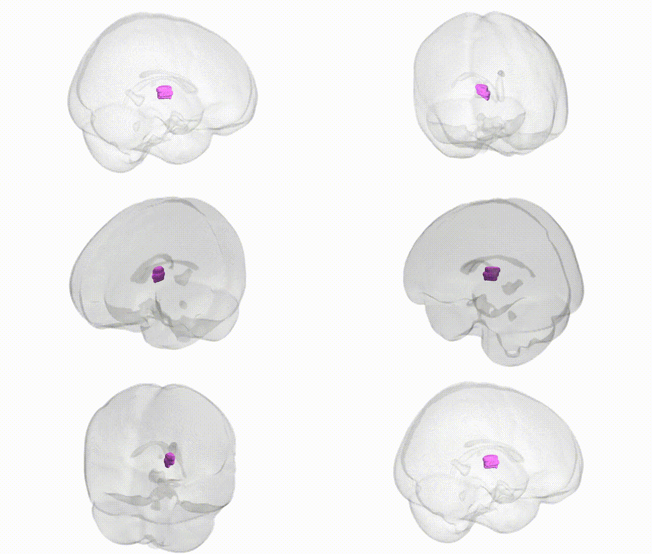
Full Quality Version: Download MP4
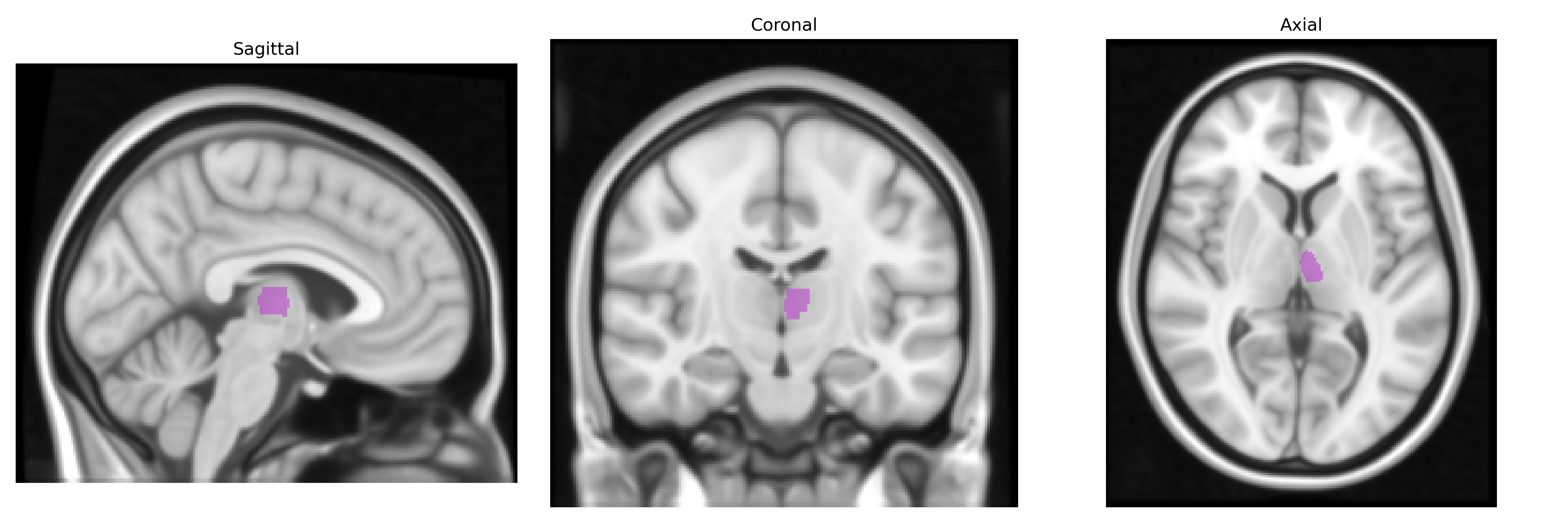
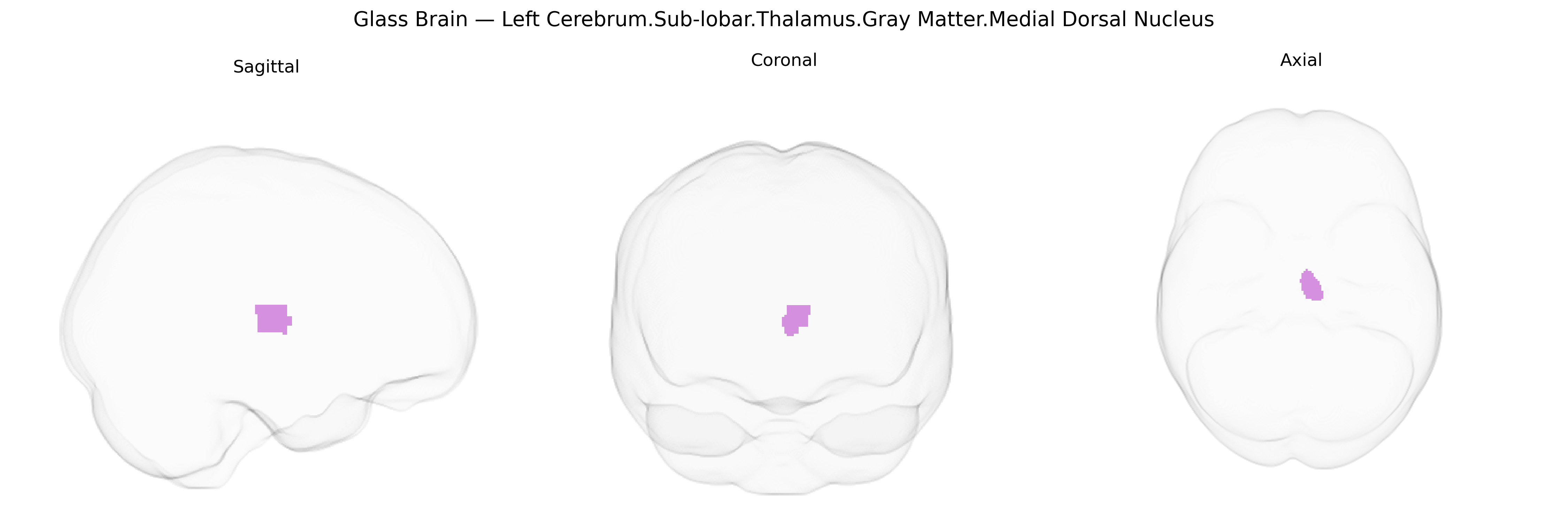
Wali Sidiqyar*, Gaurav Rudravaram*, Elyssa M. McMaster, Trent M. Schwartz, Adam M. Saunders, Kurt G. Schilling, Bennett A. Landman "Introducing SPINS: A Shared Public Visualization Library of Neuroanatomical Structures." Medical Imaging with Deep Learning- short paper
This resource is licensed under CC0 1.0 Universal (Public Domain).