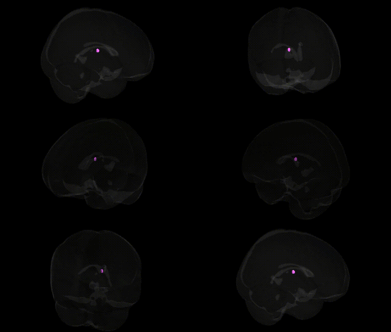
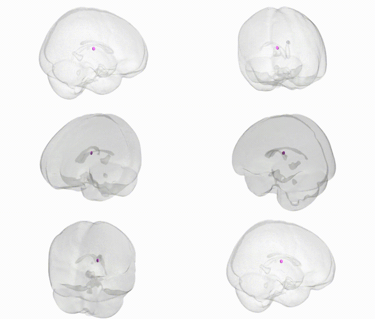
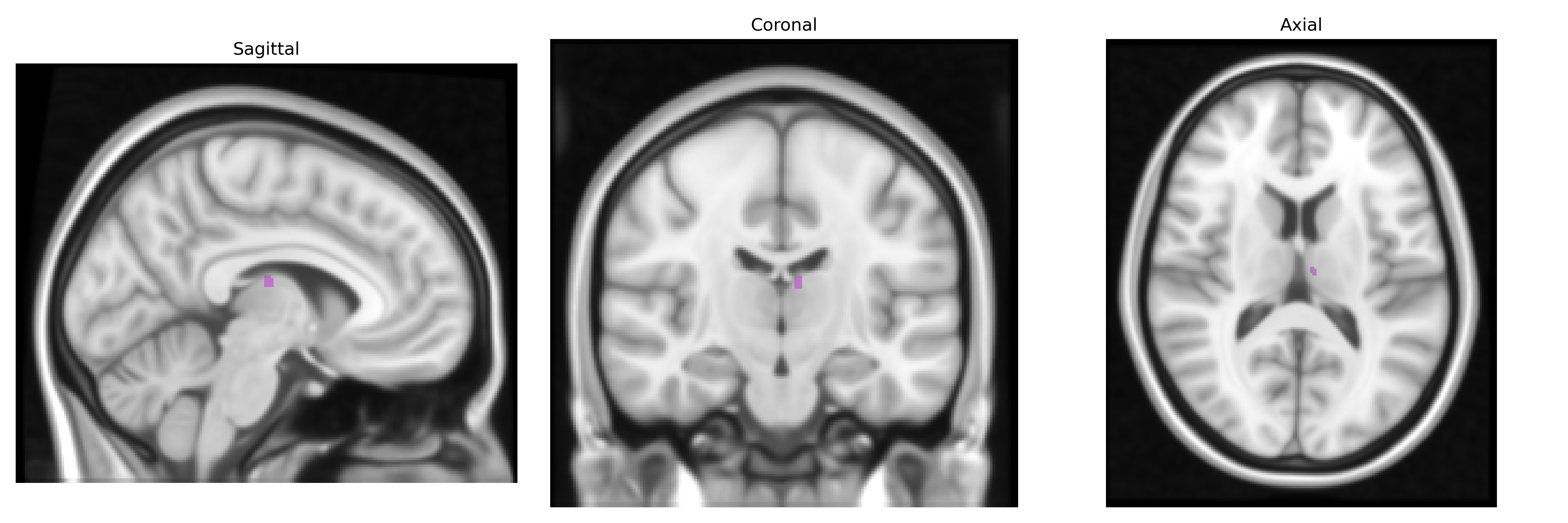
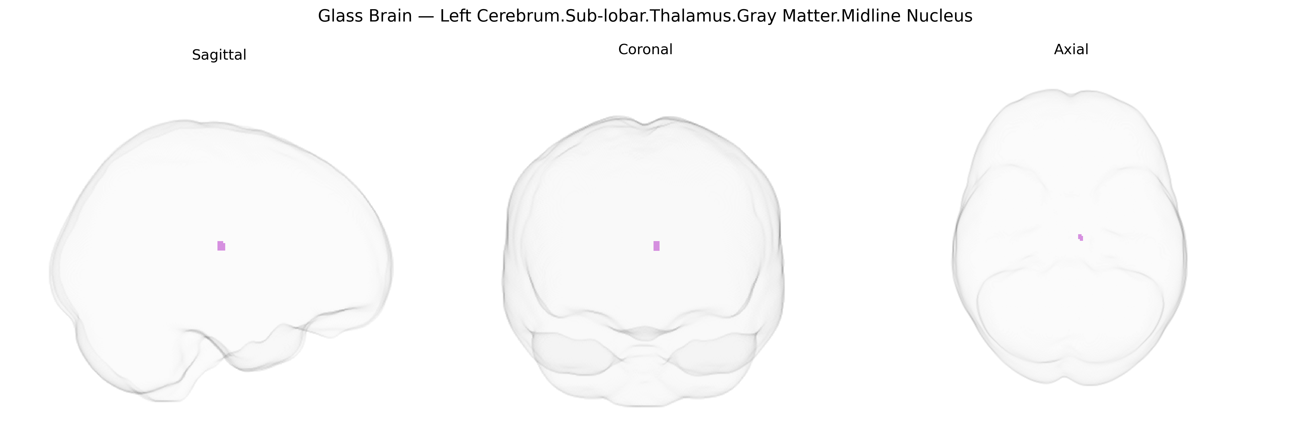
The bilateral Left Cerebrum.Sub-lobar.Thalamus.Gray Matter.Midline Nucleus corresponds to midline thalamic nuclei located along the medial thalamic wall adjacent to the third ventricle, comprising a set of small gray matter nuclei that participate in limbic, arousal, and integrative cortical–subcortical networks. These nuclei receive inputs from brainstem reticular and limbic structures and project primarily to medial prefrontal, cingulate, and other associative cortical regions, thereby contributing to regulation of consciousness, attention, emotional processing, and aspects of memory. Functionally, the midline nuclei are often grouped with intralaminar and other “nonspecific” thalamic nuclei because of their diffuse cortical projections and role in modulating global cortical excitability and synchrony. There is no direct Wikipedia article for this exact midline thalamic nuclear group; a closely related structure is the Thalamus.
Genetic associations involving the bilateral left midline thalamic nuclei (Sub-lobar Thalamus Gray Matter, midline nucleus region in the Talairach 1 mm atlas) have largely emerged from imaging-genetics and GWAS studies of thalamic volume, thalamocortical connectivity, and neuropsychiatric disorders rather than from nucleus-specific analyses, with many findings implicating polygenic, small-effect variants spread across the genome. Large-scale ENIGMA and UK Biobank–based GWAS have identified loci near genes such as MAPT, LRRC37A, DLG2, GRIN2A, and others in synaptic, glutamatergic, and neurodevelopmental pathways associated with overall thalamic volume and thalamic subregional structure, including midline and mediodorsal territories involved in limbic and cognitive circuits. Polygenic risk for schizophrenia, bipolar disorder, major depressive disorder, and autism spectrum disorder has been correlated with thalamic structural and functional alterations, with schizophrenia GWAS loci (e.g., in CACNA1C, GRIN2A, and complement pathway genes such as C4A) often cited in the context of disrupted thalamocortical signaling. Variants in genes involved in sleep–wake regulation and arousal (e.g., in CLOCK/BMAL1-related pathways and orexin/hypocretin signaling) have been linked indirectly to midline and intralaminar thalamic function through sleep and circadian GWAS, while thalamic structural GWAS have also intersected with loci implicated in neurodegenerative conditions such as Alzheimer’s disease and Parkinson’s disease (e.g., APOE, MAPT region), reflecting the vulnerability of thalamic relay and associative nuclei in these disorders. Overall, current evidence points to a distributed, pleiotropic genetic architecture influencing midline thalamic structure and function, shared across multiple psychiatric, cognitive, circadian, and neurodegenerative phenotypes, but precise gene–nucleus mappings for the specific Talairach-defined midline nucleus remain limited and are typically inferred from broader thalamic or thalamocortical GWAS signals rather than direct, nucleus-resolved genetic studies.
Overview generated by GPT-4o (2026).
Region ID: 790
Hemisphere: bilateral
Atlas: Talairach labels 1mm
Full Quality Version: Download MP4
Full Quality Version: Download MP4
Wali Sidiqyar*, Gaurav Rudravaram*, Elyssa M. McMaster, Trent M. Schwartz, Adam M. Saunders, Kurt G. Schilling, Bennett A. Landman "Introducing SPINS: A Shared Public Visualization Library of Neuroanatomical Structures." Medical Imaging with Deep Learning- short paper
This resource is licensed under CC0 1.0 Universal (Public Domain).