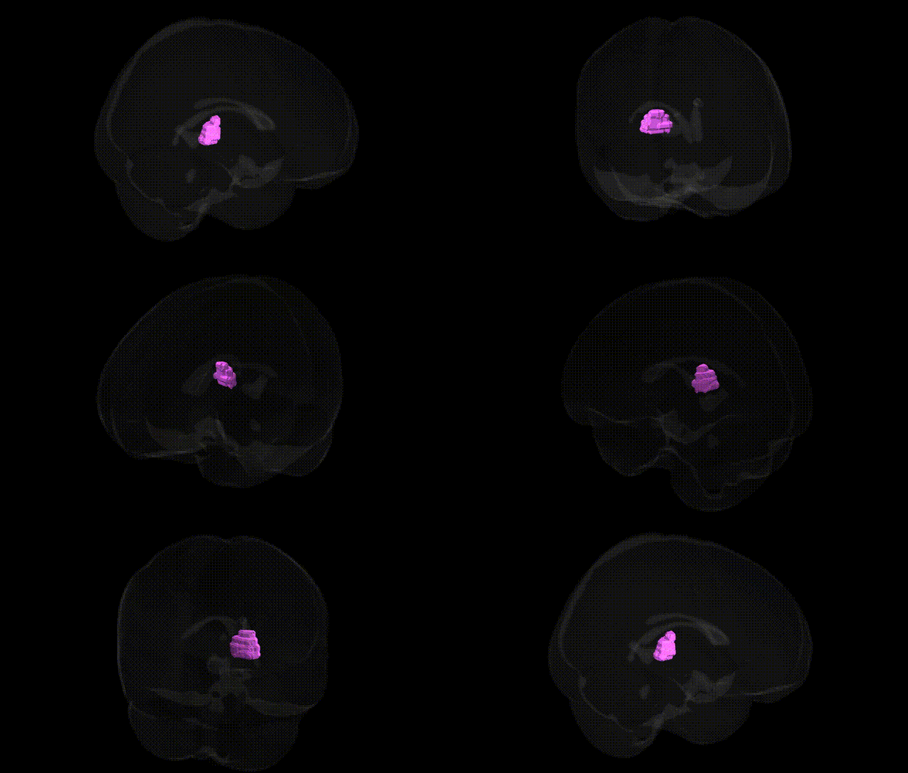
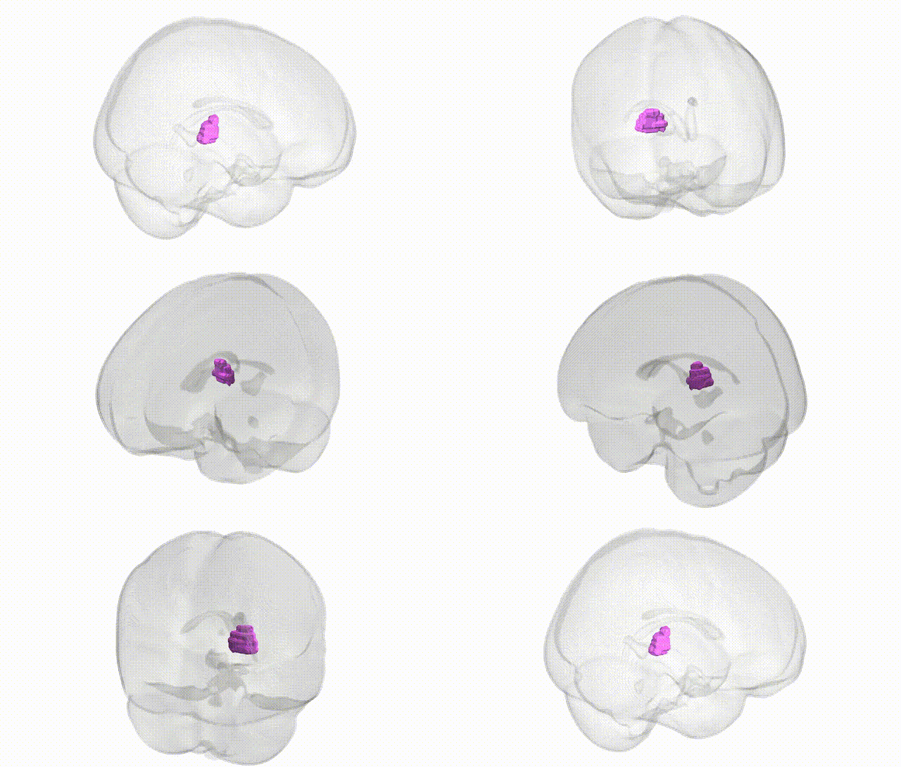
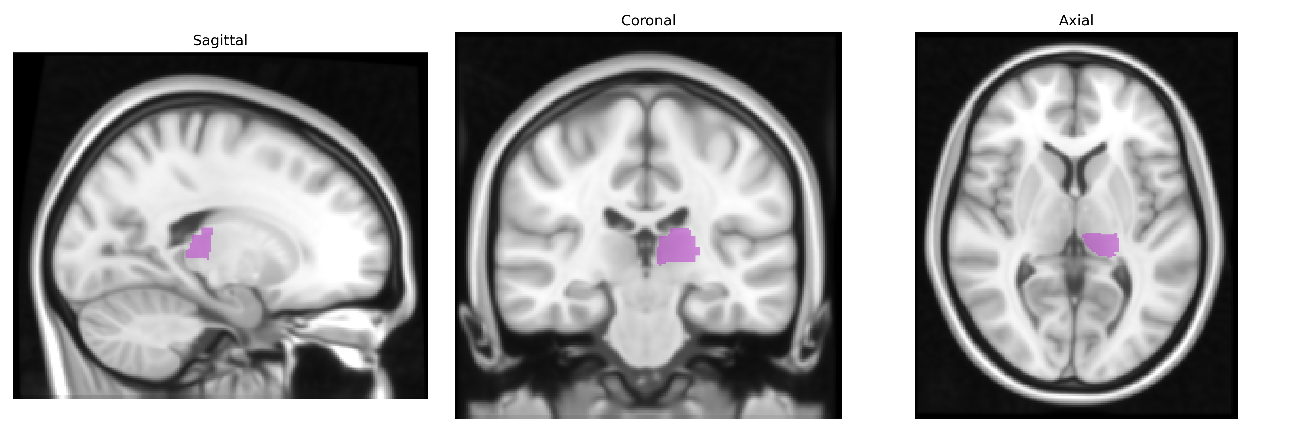
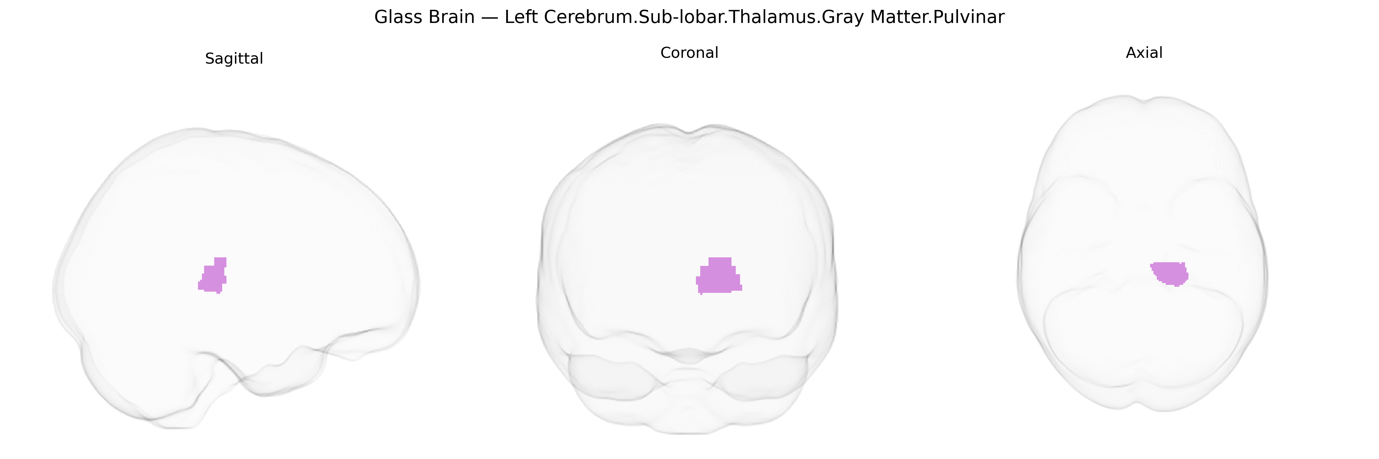
The bilateral Left Cerebrum.Sub-lobar.Thalamus.Gray Matter.Pulvinar corresponds to the pulvinar nuclei of the thalamus, a large posterior thalamic complex situated in the subcortical gray matter, forming part of the dorsal thalamus and bordering the lateral and medial geniculate bodies. It is heavily interconnected with occipital, parietal, and temporal association cortices, as well as subcortical visual and attentional networks, and is implicated in higher-order visual processing, spatial attention, and integration of multisensory information. Functionally, the pulvinar participates in filtering and gating of sensory input, modulation of cortical synchronization, and coordination of attentionally relevant signals across distributed cortical areas, thereby contributing to coherent perceptual and cognitive operations. Pulvinar
Genetic associations involving the pulvinar nucleus of the thalamus, as defined in Talairach-based parcellations (including the Left Cerebrum.Sub-lobar.Thalamus.Gray Matter.Pulvinar region), largely emerge from imaging–genetics and GWAS of subcortical volumes and thalamic nuclei rather than from locus-specific studies, and they often implicate neurodevelopmental and synaptic pathways. Large-scale GWAS of subcortical structures (for example, ENIGMA and UK Biobank analyses) have reported common variants in genes related to neurodevelopment, axon guidance, and synaptic signaling (such as variants near or within CELSR2, APOE, MAPT, and other loci) that influence thalamic volume, within which the pulvinar is a major posterior nucleus, although most work has not isolated pulvinar-specific effects. Pulvinar structural and functional alterations have been linked, via genetic or polygenic risk studies, to neuropsychiatric and neurodevelopmental disorders including schizophrenia, autism spectrum disorder, attention-deficit/hyperactivity disorder, and major depressive disorder, where risk variants in genes for glutamatergic and GABAergic signaling, calcium-channel function (e.g., CACNA1C and related loci), and synaptic plasticity show associations with thalamocortical circuitry and thalamic morphology. In movement disorders such as essential tremor and dystonia, and in some epilepsies, imaging–genetic work suggests that variants affecting thalamic network connectivity may involve the pulvinar, though these findings are typically reported at the level of thalamus or thalamic networks rather than this specific atlas label. Overall, current genetic evidence supports a role for polygenic variation influencing thalamic development and connectivity, with the pulvinar implicated as part of broader thalamocortical circuits rather than as a uniquely targeted structure with a distinct set of GWAS-identified loci.
Overview generated by GPT-4o (2026).
Region ID: 622
Hemisphere: bilateral
Atlas: Talairach labels 1mm
Full Quality Version: Download MP4
Full Quality Version: Download MP4
Wali Sidiqyar*, Gaurav Rudravaram*, Elyssa M. McMaster, Trent M. Schwartz, Adam M. Saunders, Kurt G. Schilling, Bennett A. Landman "Introducing SPINS: A Shared Public Visualization Library of Neuroanatomical Structures." Medical Imaging with Deep Learning- short paper
This resource is licensed under CC0 1.0 Universal (Public Domain).