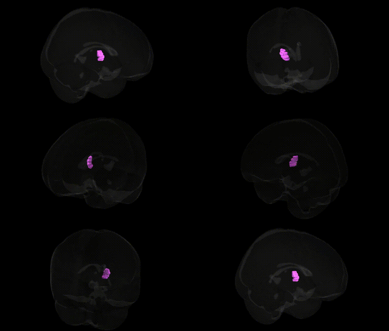
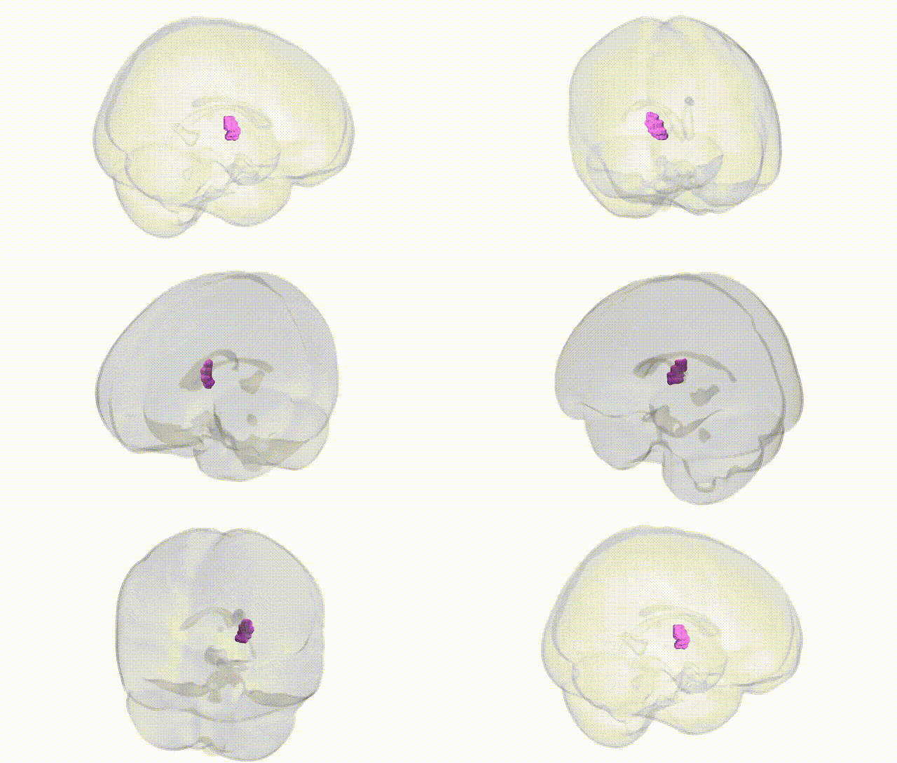
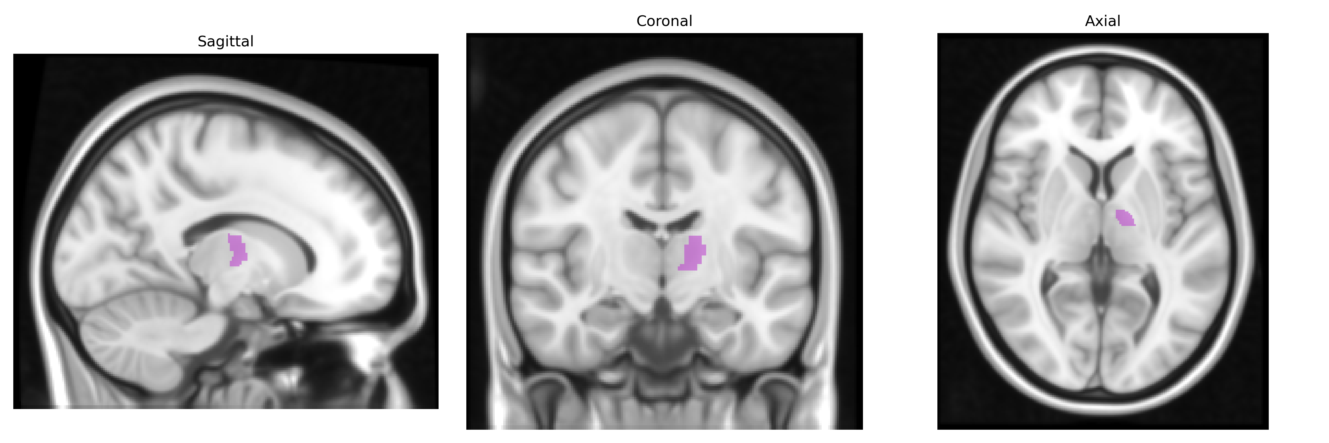
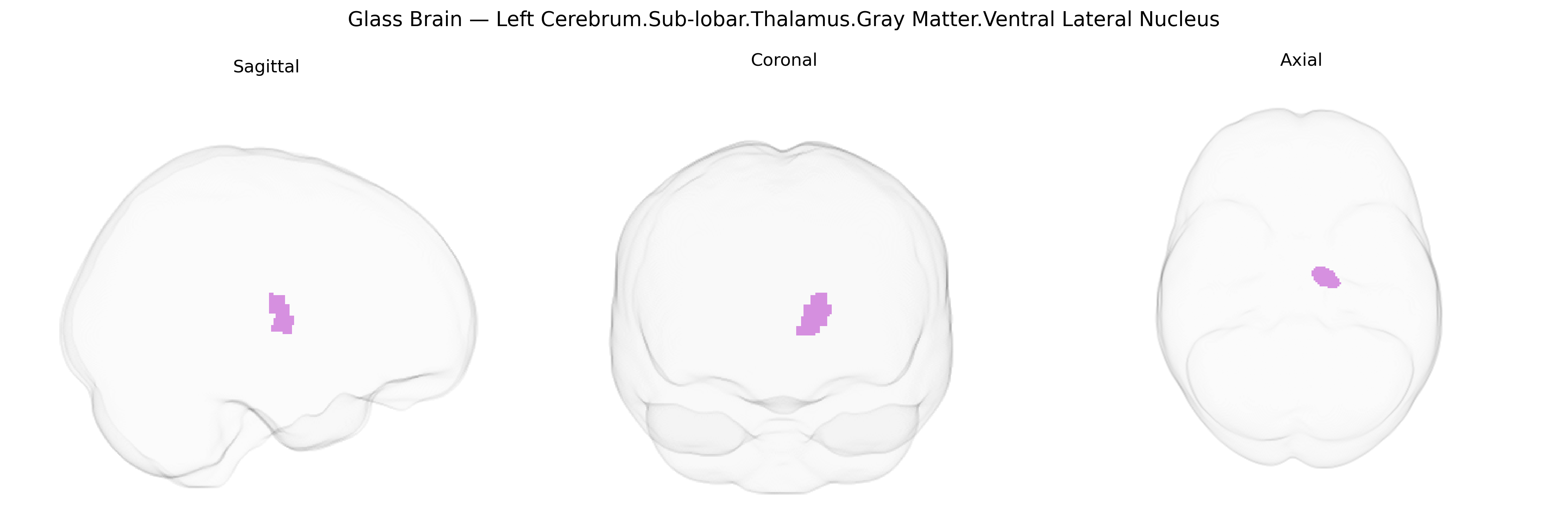
The bilateral Left Cerebrum.Sub-lobar.Thalamus.Gray Matter.Ventral Lateral Nucleus corresponds to the ventral lateral (VL) nucleus of the thalamus, a key relay and integration center within the motor thalamus. It receives dense input from the cerebellum (especially the dentate nucleus) and the globus pallidus, and projects primarily to motor and premotor cortices, thereby participating in the modulation, coordination, and planning of voluntary movement. Neurons in this nucleus help refine motor commands by integrating cerebellar signals related to movement timing and precision with basal ganglia output related to movement initiation and suppression. Functionally, the VL nucleus is implicated in motor learning, execution of fine skilled movements, and is a target or pathway node in certain movement disorders and their treatments, such as thalamic deep brain stimulation for tremor. There is no direct link for the “ventral lateral nucleus” as a standalone article; a closely related and encompassing structure is the Thalamus.
The ventral lateral (VL) nucleus of the thalamus, a sub-lobar gray-matter structure in the left cerebrum that relays cerebellar and basal ganglia signals to motor and premotor cortices, has been implicated in several genetic and genome‑wide association study (GWAS) findings through imaging genetics and disorder-focused studies, although direct VL-specific GWAS hits are rare because most analyses use broader thalamic or subcortical volumetric measures. Polygenic influences on thalamic volume and microstructure have been reported in large-scale imaging GWAS (e.g., ENIGMA, UK Biobank), with loci in or near genes involved in neurodevelopment, synaptic function, and axon guidance (such as variants in or near genes like BCL2L1, NPTX1, and others) contributing to variance in thalamic size, which presumably includes the VL nucleus. The VL thalamus shows structural and functional alterations linked to polygenic risk for movement and neuropsychiatric disorders, including Parkinson’s disease, essential tremor, dystonia, schizophrenia, bipolar disorder, and major depression, with some studies connecting thalamic changes to risk loci in genes related to dopaminergic signaling, glutamatergic transmission, and cerebello-thalamo-cortical circuitry. In epilepsy, especially generalized and absence syndromes, genetic variants influencing thalamocortical excitability and GABAergic/glutamatergic balance (e.g., in CACNA1H, GABRA genes, and others) have been associated with thalamic network dysfunction that includes the VL nucleus. Imaging–genetic work has also linked thalamic structure and connectivity, encompassing VL regions, to polygenic scores for cognitive performance, intelligence, and educational attainment, suggesting that distributed genetic influences on cortical–subcortical networks shape both VL morphology and motor–cognitive traits. Overall, the genetic architecture of the ventral lateral thalamus appears to be highly polygenic and shared with broader thalamic and motor-circuit phenotypes, with disorder-associated loci impacting this region mainly through effects on neurodevelopmental pathways and synaptic and circuit-level function rather than through nucleus-specific variants identified to date.
Overview generated by GPT-4o (2026).
Region ID: 643
Hemisphere: bilateral
Atlas: Talairach labels 1mm
Full Quality Version: Download MP4
Full Quality Version: Download MP4
Wali Sidiqyar*, Gaurav Rudravaram*, Elyssa M. McMaster, Trent M. Schwartz, Adam M. Saunders, Kurt G. Schilling, Bennett A. Landman "Introducing SPINS: A Shared Public Visualization Library of Neuroanatomical Structures." Medical Imaging with Deep Learning- short paper
This resource is licensed under CC0 1.0 Universal (Public Domain).