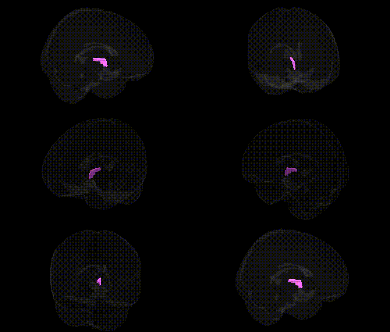
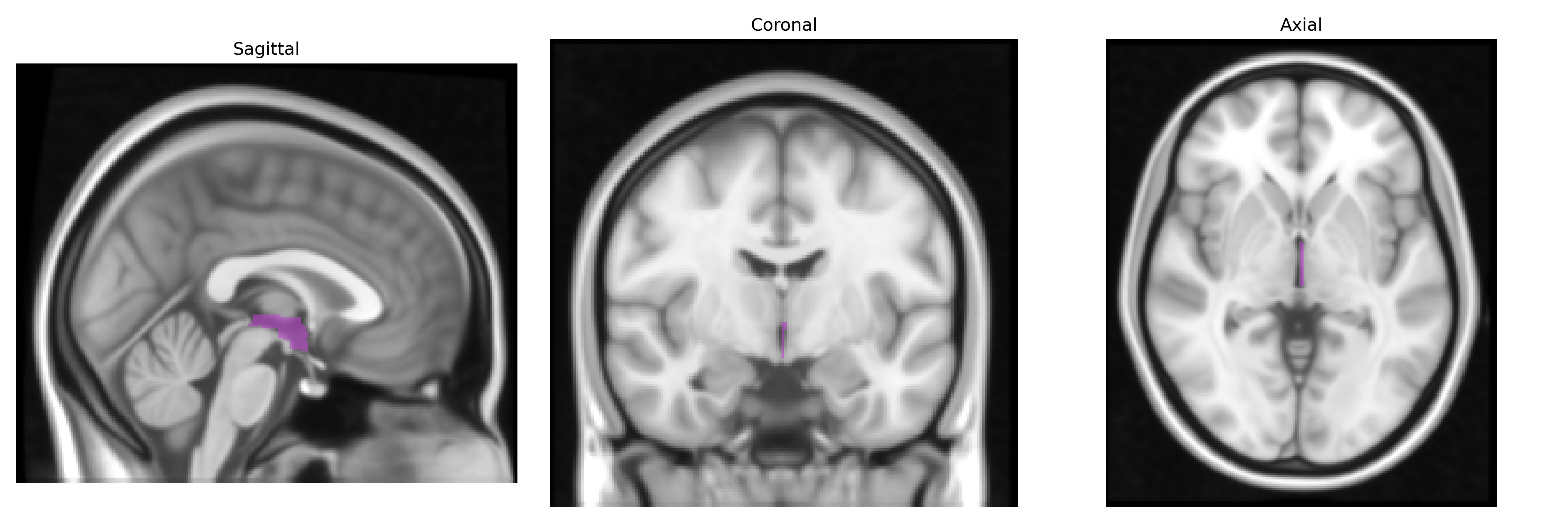
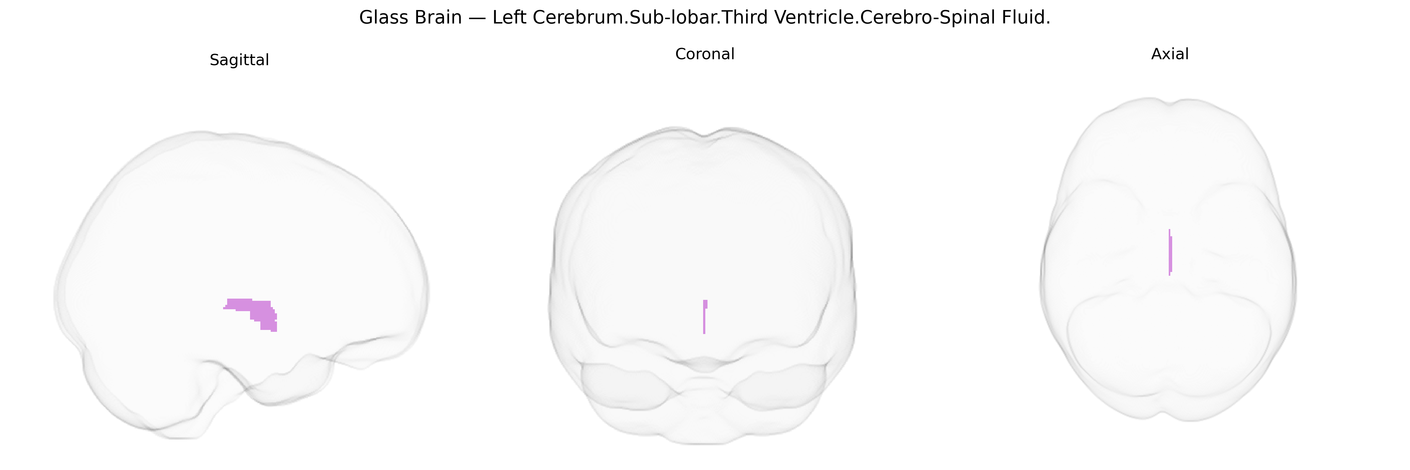
The bilateral Left Cerebrum.Sub-lobar.Third Ventricle.Cerebro-Spinal Fluid region corresponds to the cerebrospinal fluid (CSF)–filled cavity of the third ventricle located in the midline diencephalon, bordered primarily by the thalamus and hypothalamus. It forms part of the ventricular system, connecting the lateral ventricles via the interventricular foramina (foramina of Monro) anteriorly and the cerebral aqueduct posteriorly, thereby contributing to CSF circulation between supratentorial and infratentorial compartments. CSF within the third ventricle cushions neural tissue, helps maintain intracranial pressure, and participates in metabolic waste clearance and neurochemical homeostasis. The Talairach label “Sub-lobar” reflects its deep, central location beneath cortical lobes rather than a specific functional nucleus. There is no direct link for this exact compound label; a related structure is the Third Ventricle.
The third ventricle, a midline cerebrospinal fluid (CSF) space within the sub-lobar region of the left cerebrum, is frequently examined in genetic imaging studies as a proxy for global and subcortical atrophy rather than as a functionally specific “region,” and genome-wide association studies (GWAS) have identified multiple loci associated with its volume and related CSF measures. Polygenic influences on third ventricle and overall ventricular volume include variants in or near genes implicated in neurodevelopment, myelination, and neurodegeneration—such as APOE (particularly ε4), MAPT, and other Alzheimer’s disease–related loci—reflecting shared genetic architecture with cortical and hippocampal atrophy, white matter loss, and dementia risk. Enlarged third ventricle volume shows heritability and has been genetically correlated with schizophrenia, bipolar disorder, and major depressive disorder, consistent with broader ventricular enlargement phenotypes observed in psychiatric GWAS and imaging–genetics consortia (e.g., ENIGMA). Additional associations have been reported with genes involved in CSF dynamics and ependymal or choroid plexus function, though these are less well replicated, and with polygenic scores for neurodevelopmental and cognitive traits that influence overall brain structure. Overall, genetic findings linked to the Talairach-defined left sub-lobar third ventricle CSF region primarily reflect genes affecting global brain atrophy, neurodegenerative disease susceptibility, and major psychiatric disorders through their impact on ventricular enlargement rather than region-specific molecular mechanisms.
Overview generated by GPT-4o (2026).
Region ID: 362
Hemisphere: bilateral
Atlas: Talairach labels 1mm
Full Quality Version: Download MP4
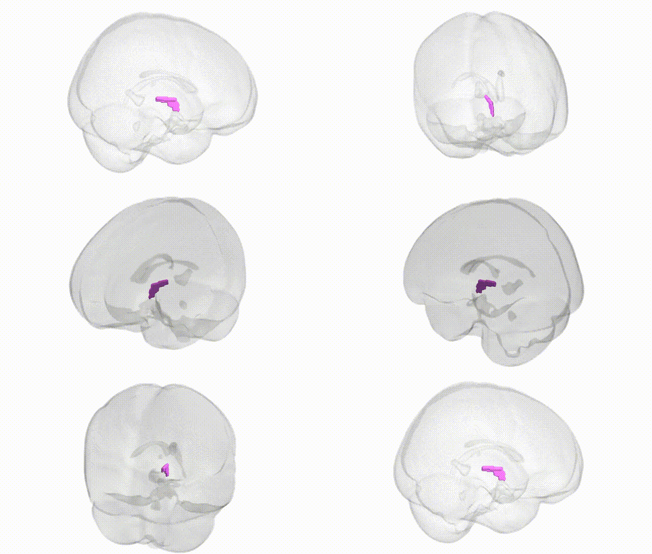
Full Quality Version: Download MP4
Wali Sidiqyar*, Gaurav Rudravaram*, Elyssa M. McMaster, Trent M. Schwartz, Adam M. Saunders, Kurt G. Schilling, Bennett A. Landman "Introducing SPINS: A Shared Public Visualization Library of Neuroanatomical Structures." Medical Imaging with Deep Learning- short paper
This resource is licensed under CC0 1.0 Universal (Public Domain).