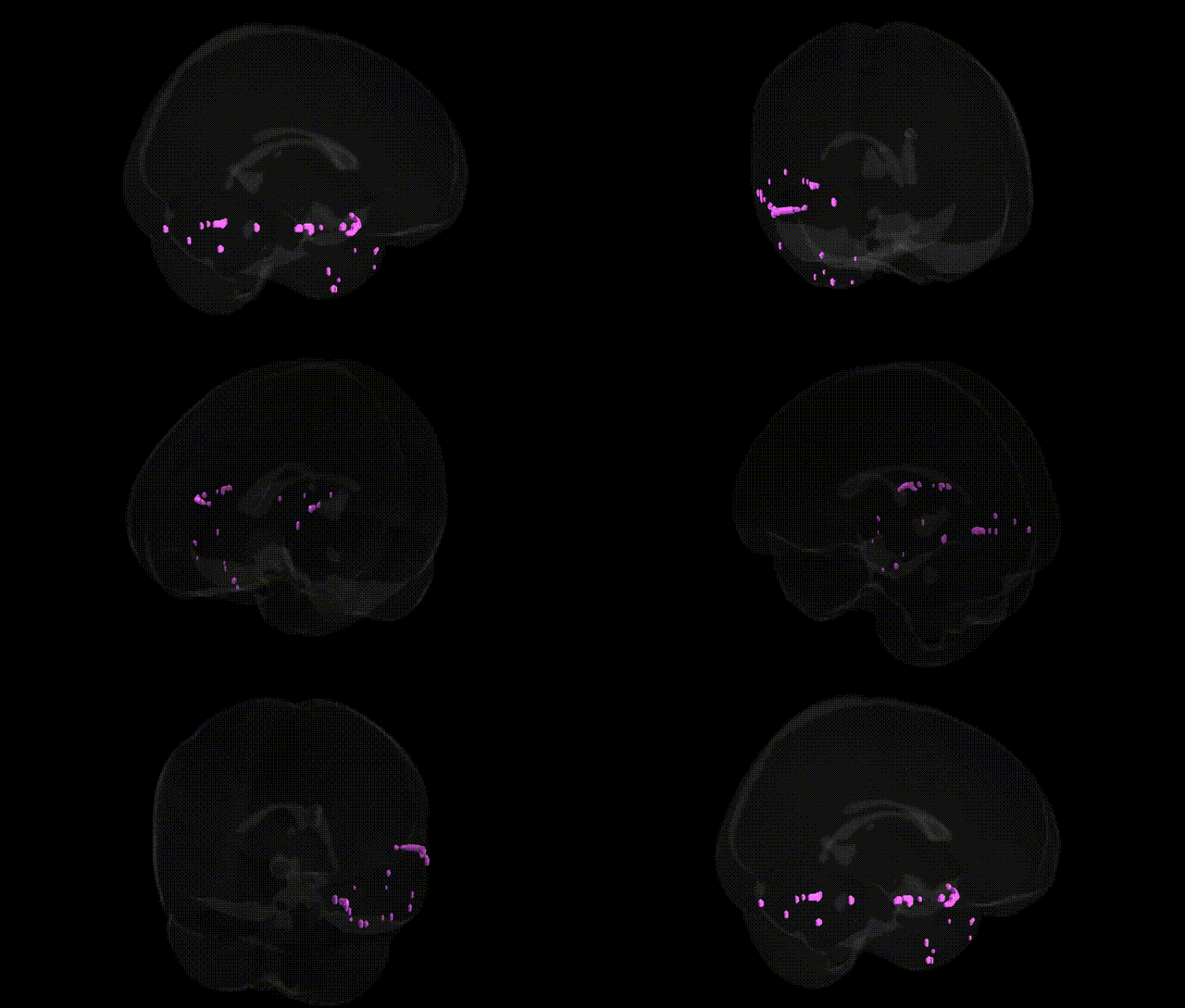
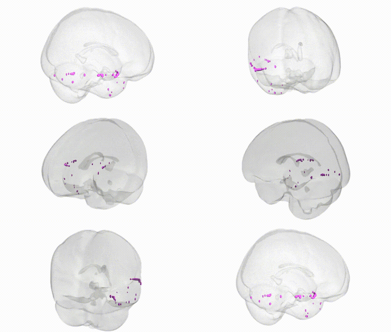
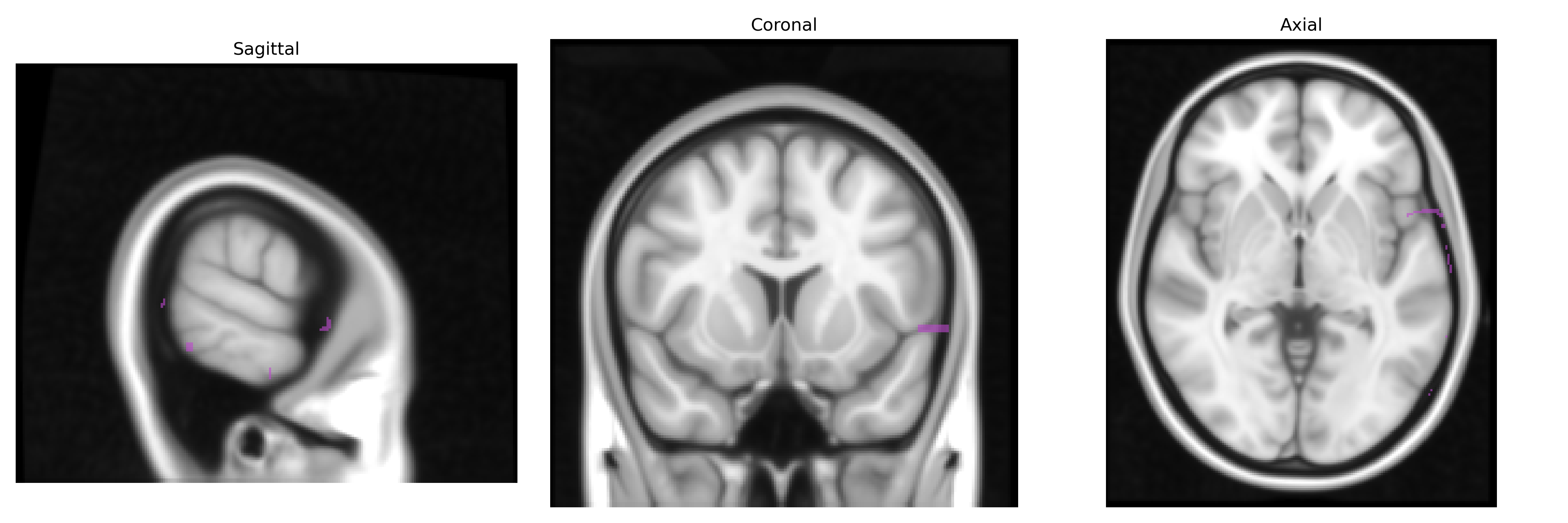
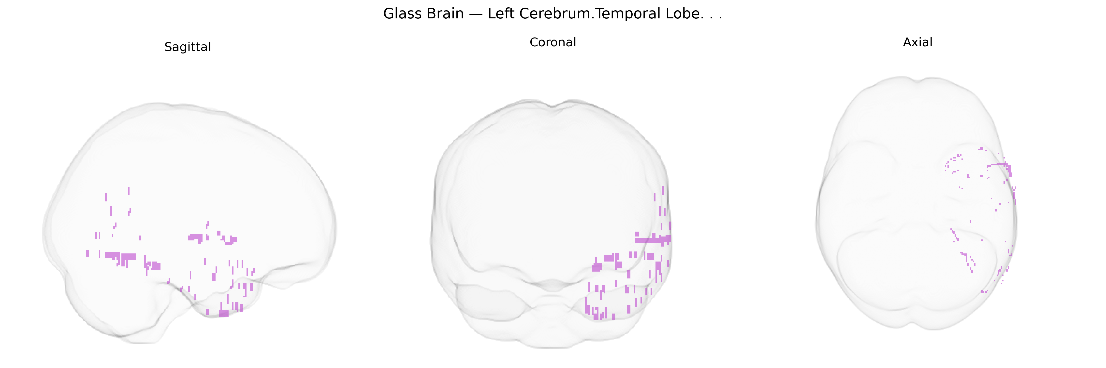
The bilateral left cerebrum temporal lobe is a paired cortical region located inferior to the lateral (Sylvian) fissure and anterior to the occipital lobe, encompassing lateral, superior, middle, and inferior temporal gyri as well as medial structures such as parts of the hippocampus and amygdala. It plays a central role in auditory processing, language comprehension (particularly in the dominant hemisphere, typically left), semantic memory, and aspects of visual object recognition and social cognition. The temporal lobe also contributes critically to episodic memory encoding and retrieval via medial temporal structures, integrates multimodal sensory inputs, and supports higher-order processes such as emotion, music perception, and recognition of complex sounds and faces. A related article is Temporal lobe.
The bilateral left temporal lobe (including superior, middle, and inferior temporal gyri as defined in Talairach 1 mm space) is strongly implicated in genetic studies of language, auditory processing, and higher-order cognition, with multiple GWAS, imaging-genetics, and candidate-gene studies linking variation in this region’s structure and function to specific loci and pathways. Common variants in FOXP2, CNTNAP2, DCDC2, KIAA0319, ROBO1, and other neurodevelopmental genes have been associated with altered left temporal morphology and language-related activation patterns, particularly in superior and middle temporal regions involved in phonological and semantic processing. Large-scale neuroimaging GWAS (e.g., ENIGMA, UK Biobank) have identified numerous loci (such as in genes like HMGA2, TESC, and others affecting neurogenesis, cortical patterning, and synaptic function) that influence temporal lobe cortical thickness, surface area, and gyrification, traits that show significant heritability. Left temporal lobe anomalies are genetically linked to primary progressive aphasia and temporal-variant frontotemporal dementia, with mutations in GRN, MAPT, and C9orf72 contributing to selective temporal degeneration, while temporal lobe epilepsy has been associated with variants in genes affecting excitability and synaptic transmission (e.g., SCN1A and related ion channel genes). Schizophrenia, bipolar disorder, major depression, and autism spectrum disorders show consistent temporal lobe structural and functional alterations, and polygenic risk scores for these conditions correlate with variation in left temporal volume and connectivity, implicating synaptic, immune (e.g., complement system), and neurodevelopmental pathways. Overall, the bilateral left temporal lobe emerges as a key neuroanatomical substrate in genetic architectures of language, epilepsy, neurodegeneration, and major psychiatric disorders, with many implicated loci exerting broad effects on cortical development and network organization rather than being specific to this region alone.
Overview generated by GPT-4o (2026).
Region ID: 10
Hemisphere: bilateral
Atlas: Talairach labels 1mm
Full Quality Version: Download MP4
Full Quality Version: Download MP4
Wali Sidiqyar*, Gaurav Rudravaram*, Elyssa M. McMaster, Trent M. Schwartz, Adam M. Saunders, Kurt G. Schilling, Bennett A. Landman "Introducing SPINS: A Shared Public Visualization Library of Neuroanatomical Structures." Medical Imaging with Deep Learning- short paper
This resource is licensed under CC0 1.0 Universal (Public Domain).