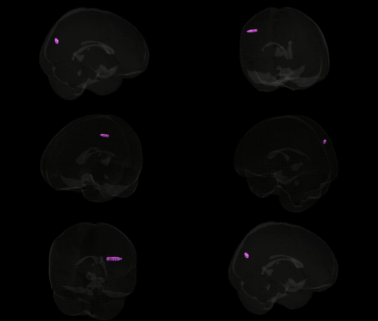
The bilateral left cerebrum temporal lobe angular gyrus white matter refers to the subcortical fiber pathways underlying the angular gyrus, a multimodal association region located at the junction of the parietal, temporal, and occipital lobes in the posterior part of the superior temporal and inferior parietal region. This white matter contains long association fibers (including components of the superior longitudinal fasciculus and other temporo-parietal tracts) that interconnect auditory, visual, and somatosensory association cortices, supporting functions such as language comprehension, reading and writing, number processing, spatial cognition, and aspects of semantic memory. Disruption of these white matter pathways, for example by stroke or degenerative disease, can contribute to classic angular gyrus syndromes, including elements of Gerstmann’s syndrome and various aphasic and alexic manifestations. There is no direct link for the white matter component alone; a related cortical region is the Angular gyrus.
The angular gyrus white matter in the left temporal–parietal region (often labeled in Talairach-based atlases as Left Cerebrum.Temporal Lobe.Angular Gyrus.White Matter) is strongly implicated by genetic and imaging–genetics studies in language, reading, and higher cognitive functions, with multiple genome-wide association studies (GWAS) linking common variants to its structure and connectivity. Variants in genes involved in axonal guidance, synaptic plasticity, and myelination—such as those in DCDC2, KIAA0319, ROBO1, CNTNAP2, and FOXP2-associated networks—have been repeatedly associated with reading ability, dyslexia, and broader language-related traits, where structural and diffusion MRI show altered white-matter microstructure in the left temporo‑parietal/angular gyrus region. Large-scale GWAS of cortical surface area, thickness, and white-matter integrity (e.g., ENIGMA consortia and UK Biobank) have identified polygenic influences on temporo‑parietal white matter, with enrichment in neurodevelopmental pathways and genes regulating neuronal migration and oligodendrocyte function, although associations are usually reported for broader temporal or parietal regions rather than the angular gyrus label specifically. Additionally, polygenic risk for schizophrenia, autism spectrum disorder, and attention-deficit/hyperactivity disorder has been associated with altered connectivity and microstructure in left temporo‑parietal white matter tracts (including the superior longitudinal fasciculus and arcuate fasciculus that traverse the angular gyrus), while Alzheimer’s disease and other dementias show GWAS-linked risk loci (e.g., APOE, CLU, PICALM) associated with white-matter degeneration and hypoconnectivity in posterior temporo‑parietal networks that include the angular gyrus, collectively indicating that the genetic architecture of this region’s white matter involves overlapping neurodevelopmental, neuropsychiatric, and neurodegenerative pathways.
Overview generated by GPT-4o (2026).
Region ID: 961
Hemisphere: bilateral
Atlas: Talairach labels 1mm
Full Quality Version: Download MP4
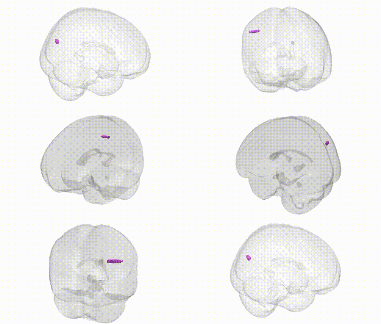
Full Quality Version: Download MP4
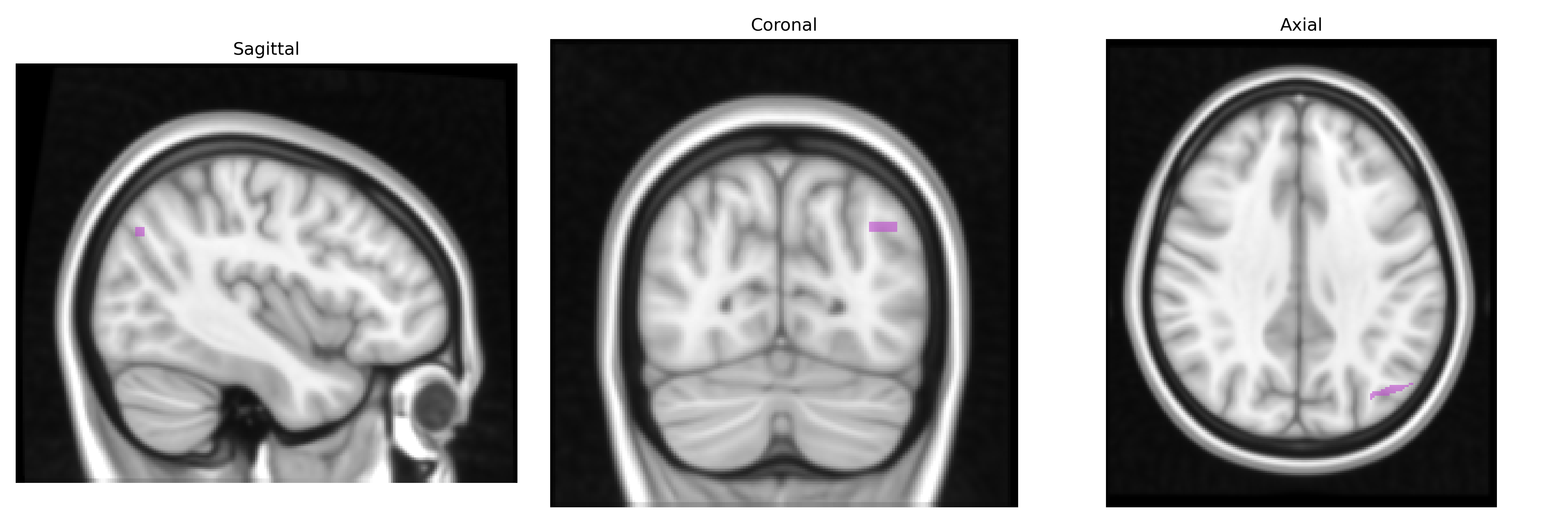
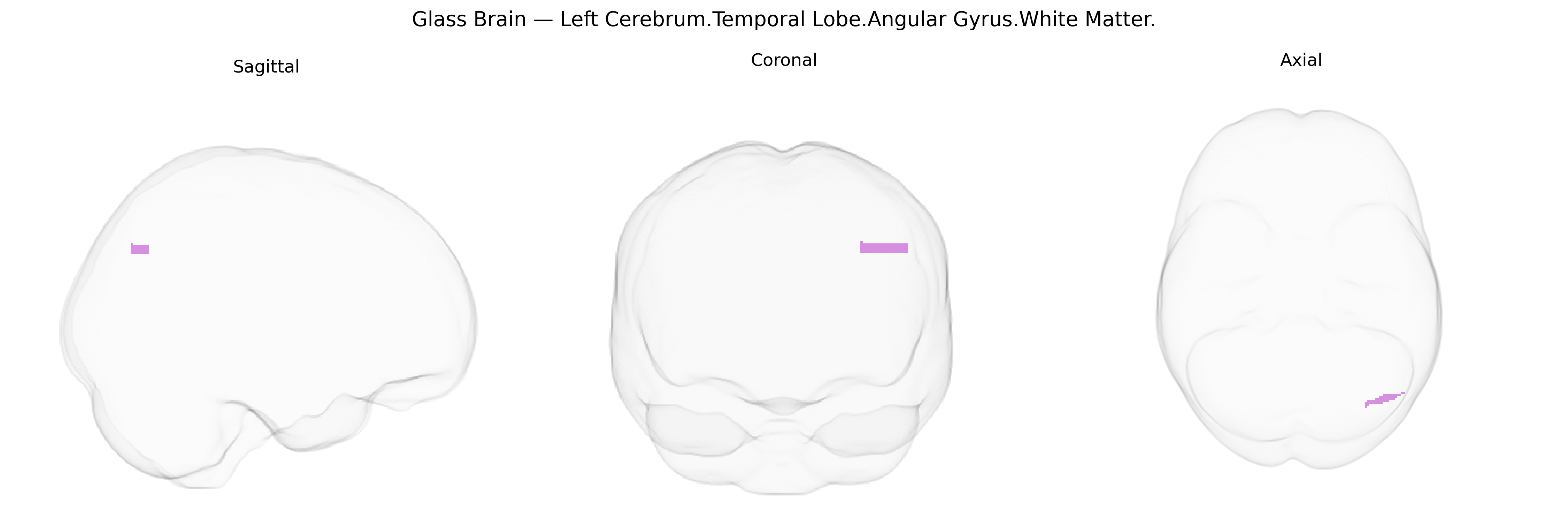
Wali Sidiqyar*, Gaurav Rudravaram*, Elyssa M. McMaster, Trent M. Schwartz, Adam M. Saunders, Kurt G. Schilling, Bennett A. Landman "Introducing SPINS: A Shared Public Visualization Library of Neuroanatomical Structures." Medical Imaging with Deep Learning- short paper
This resource is licensed under CC0 1.0 Universal (Public Domain).