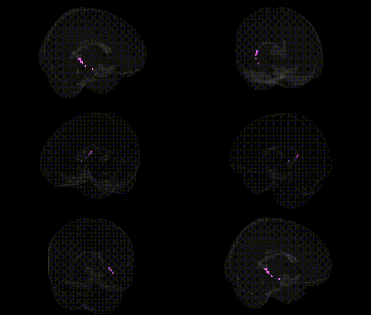
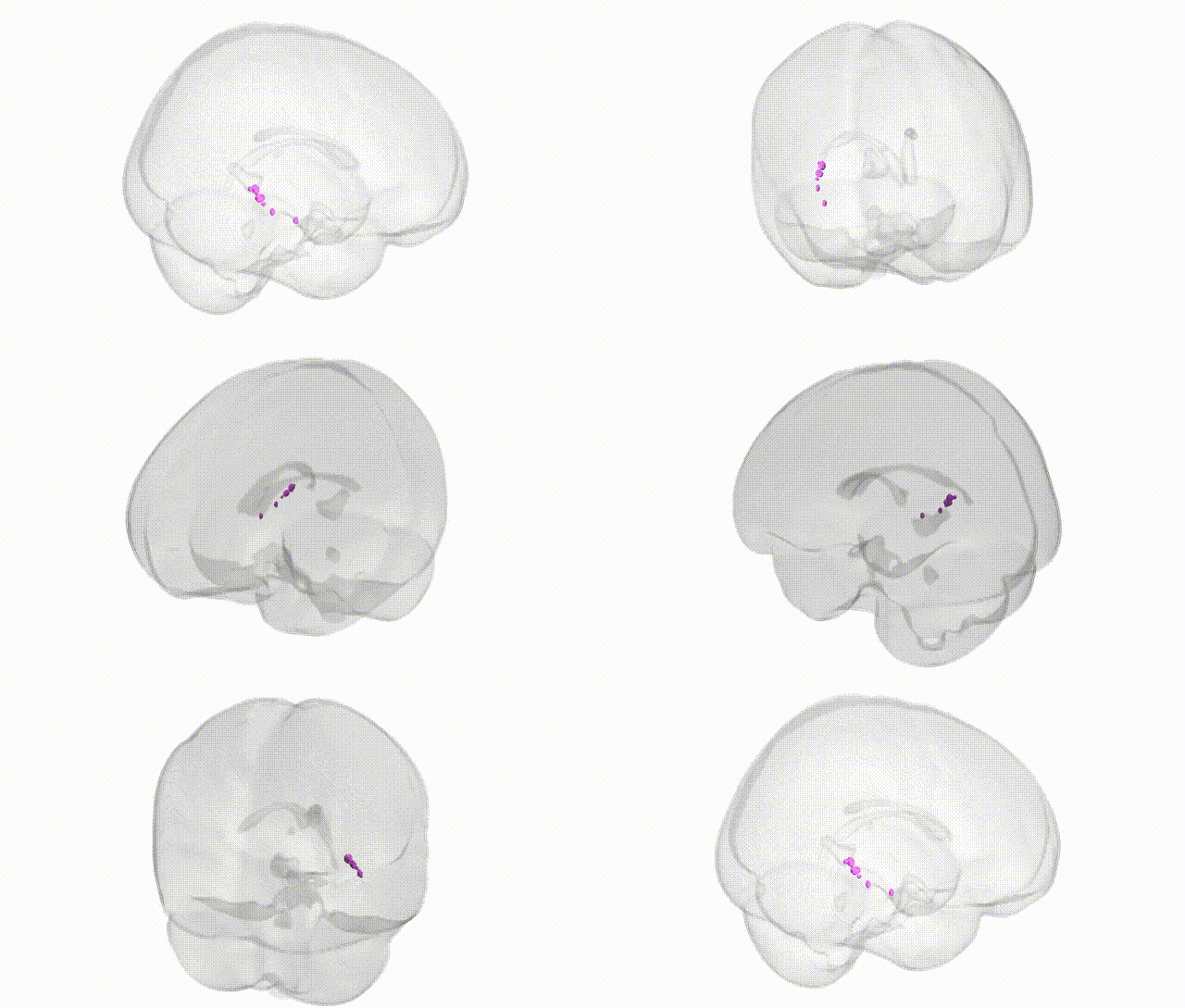
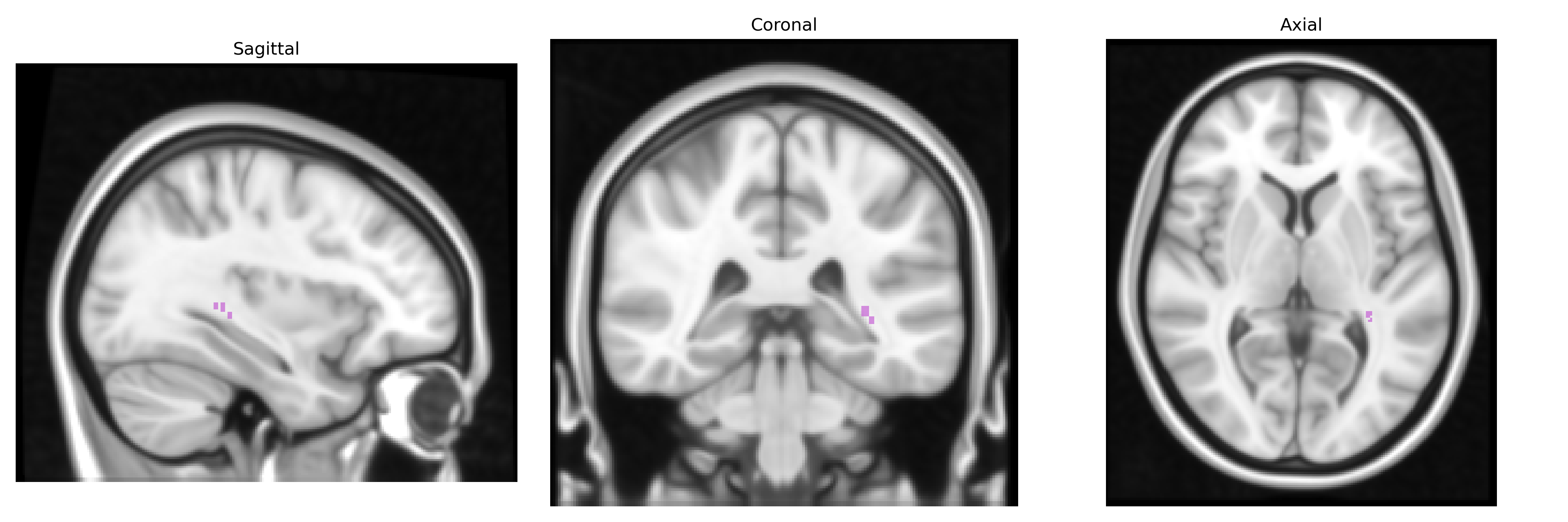
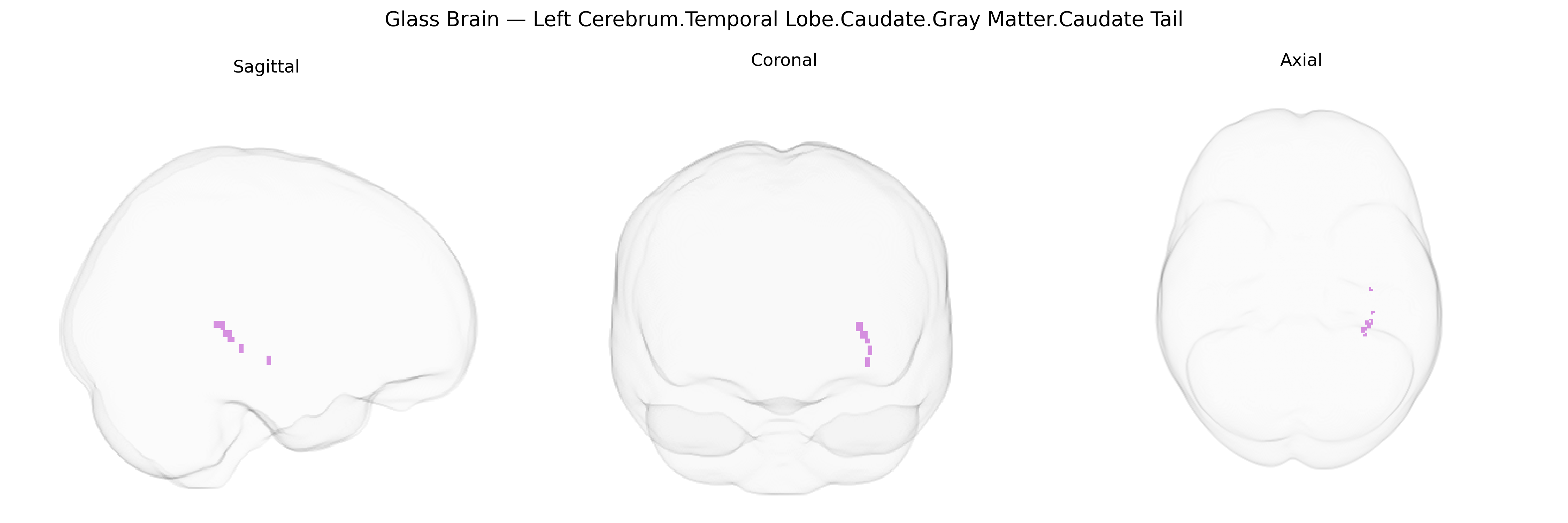
The bilateral Left Cerebrum.Temporal Lobe.Caudate.Gray Matter.Caudate Tail region corresponds to the tail portion of the caudate nucleus, a C-shaped gray matter structure that arches posteriorly from the body of the caudate along the temporal horn of the lateral ventricle into the temporal lobe. The caudate tail is part of the dorsal striatum within the basal ganglia and is heavily interconnected with association cortices, particularly visual and temporal regions, contributing to functions such as visuospatial processing, habit formation, and certain aspects of procedural learning and cognitive control. Although the tail is less frequently distinguished functionally from the head and body in human studies, it shares the caudate’s general roles in action selection, reward-related processing, and modulation of cortical activity via basal ganglia–thalamocortical loops. There is no direct Wikipedia article for the caudate tail; a related structure is the Caudate nucleus.
Genetic associations involving the caudate tail region of the temporal lobe, as approximated by the Talairach 1 mm atlas label “bilateral Left Cerebrum.Temporal Lobe.Caudate.Gray Matter.Caudate Tail,” are typically inferred from studies of the caudate nucleus and temporostriatal circuitry rather than this subregion alone. Large-scale GWAS of subcortical volumes (e.g., ENIGMA and UK Biobank cohorts) have identified common variants in and near genes such as HMGA2, FAT3, IGF1, PTCH1, and others that influence caudate volume, with some signals overlapping loci also implicated in neurodevelopmental traits and general brain size. Polygenic risk for disorders including schizophrenia, bipolar disorder, ADHD, and major depressive disorder shows associations with caudate volume and connectivity, suggesting shared genetic architecture between psychiatric risk loci and structural variation in this region. Candidate gene and imaging-genetics studies have further linked dopaminergic (e.g., DRD2, COMT) and glutamatergic or synaptic genes (e.g., GRIN2B, BDNF) to caudate structure and function, consistent with the role of the tail of the caudate in habit learning, reward prediction, and corticostriatal integration. Additionally, genetic risk loci for Parkinson’s disease and other movement disorders (e.g., LRRK2, SNCA) are associated with broader basal ganglia circuitry that includes the caudate, and GWAS of cognitive traits and educational attainment frequently implicate loci that also modulate subcortical morphology, providing indirect evidence that genetic variation influencing the caudate tail contributes to individual differences in cognition and vulnerability to neuropsychiatric disease.
Overview generated by GPT-4o (2026).
Region ID: 351
Hemisphere: bilateral
Atlas: Talairach labels 1mm
Full Quality Version: Download MP4
Full Quality Version: Download MP4
Wali Sidiqyar*, Gaurav Rudravaram*, Elyssa M. McMaster, Trent M. Schwartz, Adam M. Saunders, Kurt G. Schilling, Bennett A. Landman "Introducing SPINS: A Shared Public Visualization Library of Neuroanatomical Structures." Medical Imaging with Deep Learning- short paper
This resource is licensed under CC0 1.0 Universal (Public Domain).