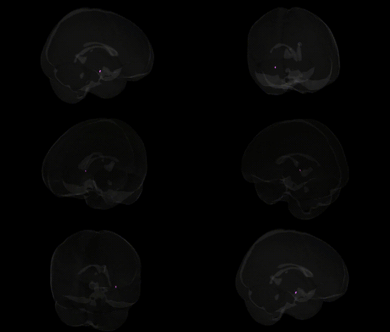
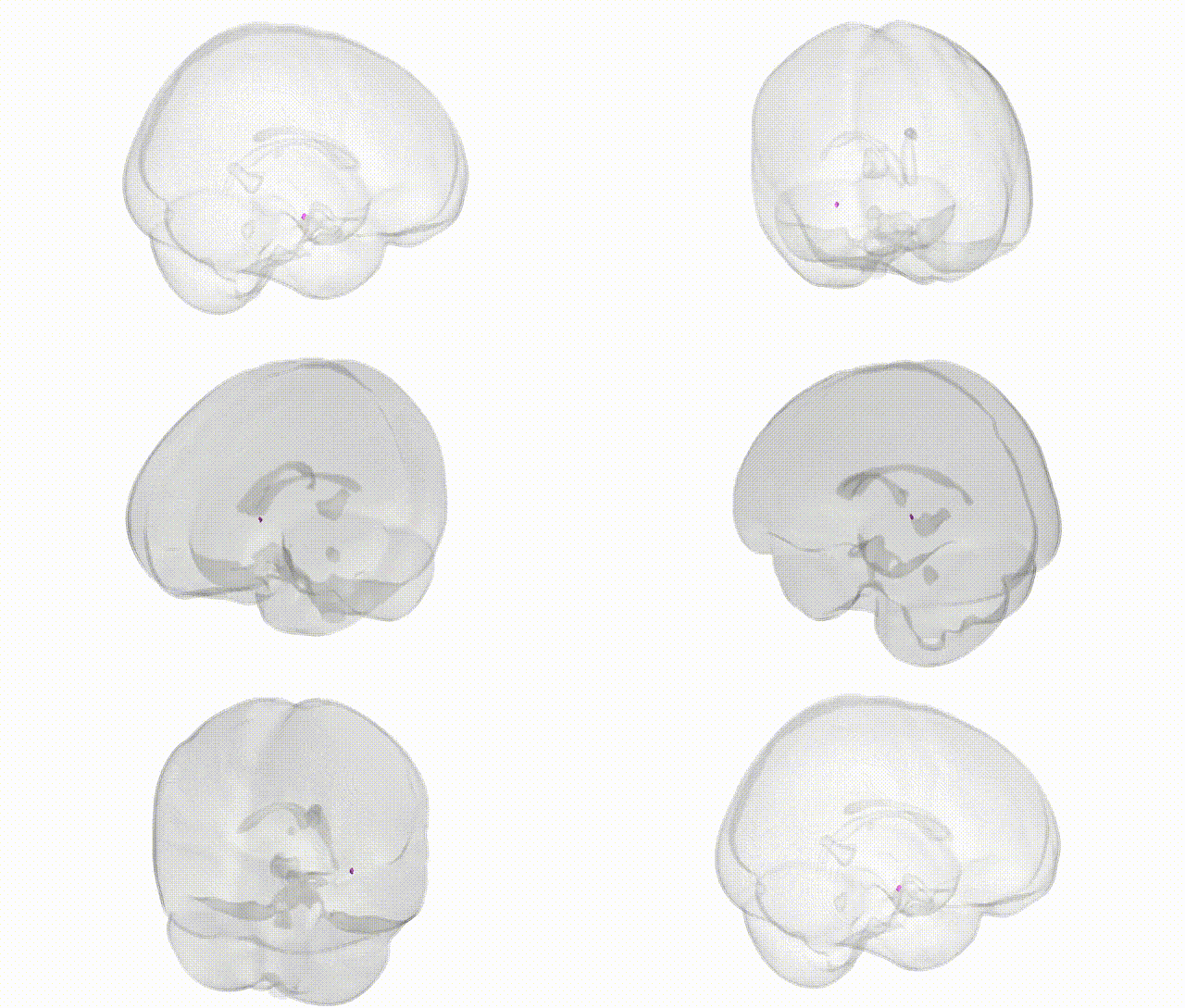
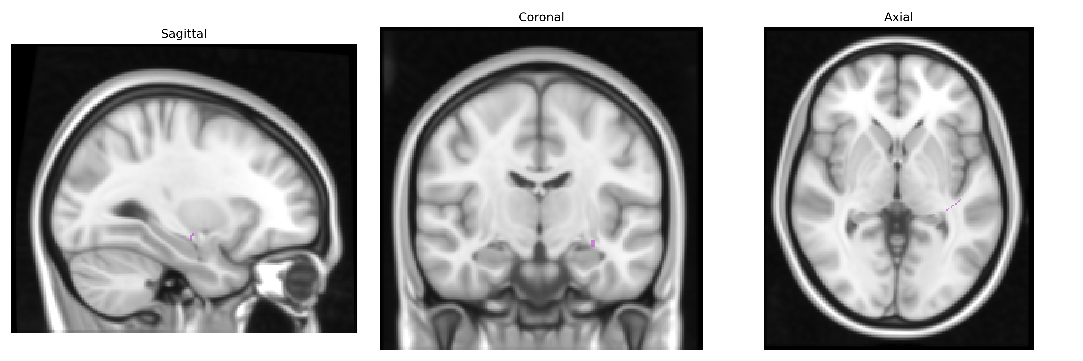
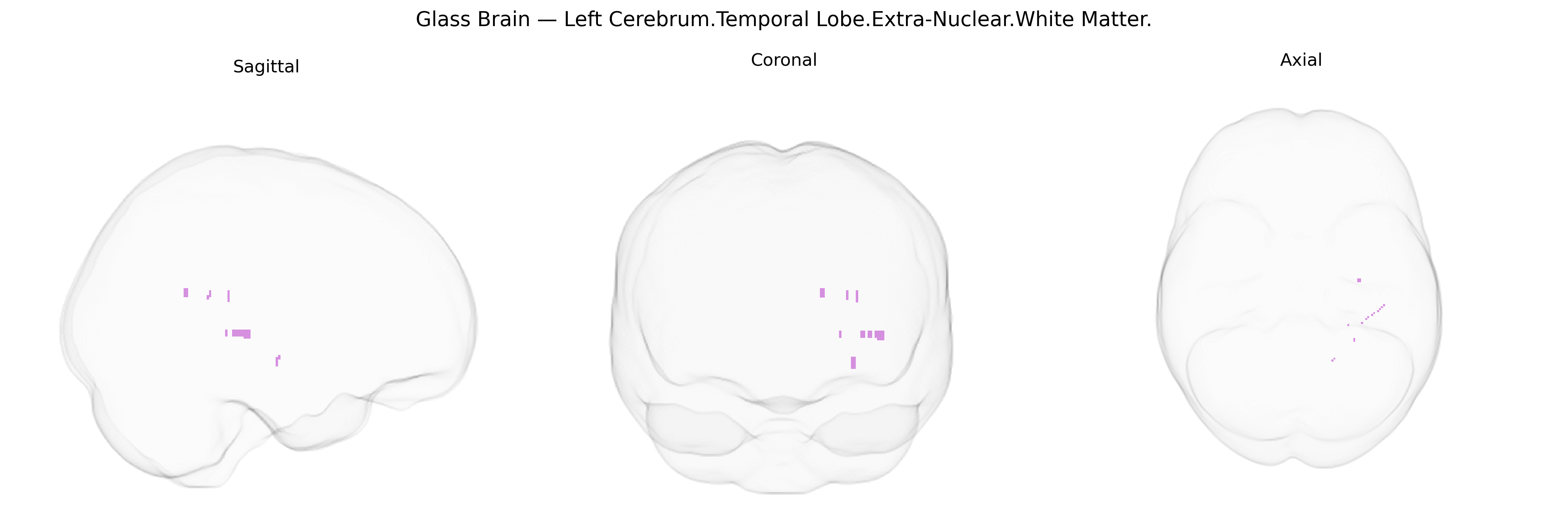
The bilateral Left Cerebrum.Temporal Lobe.Extra-Nuclear.White Matter corresponds to subcortical temporal lobe white matter tracts lying outside the major nuclear gray matter structures (such as the basal ganglia and thalamus). These regions contain long-range myelinated fibers interconnecting temporal cortical areas with frontal, parietal, occipital, and limbic regions, including components of association pathways like the inferior longitudinal fasciculus and uncinate fasciculus. Functionally, these tracts support higher-order auditory processing, language, semantic memory, and aspects of emotion and social cognition by enabling efficient communication between temporal neocortex, medial temporal structures, and distributed cortical networks. There is no direct link for this specific Talairach label; a related structure is the Temporal lobe.
Genetic associations involving bilateral left temporal lobe extra-nuclear white matter (as approximated by Talairach 1 mm atlas labels) largely emerge from imaging–genetics and GWAS of white matter microstructure and temporal lobe connectivity rather than from that specific parcel alone; common variants in genes such as BDNF, NTRK2, and COMT have been linked to individual differences in temporal lobe white matter integrity, while large-scale diffusion MRI GWAS (e.g., UK Biobank–based studies) implicate numerous loci involved in axon guidance, myelination, and oligodendrocyte function (including variants near or in genes like MAG, MBP, and CNTN4) that affect temporal association pathways. Polygenic risk for schizophrenia, bipolar disorder, and major depressive disorder is associated with altered temporal white matter metrics, and disorder-specific GWAS highlight genes related to synaptic plasticity and neurodevelopment that show downstream effects on temporal lobe tracts; similarly, autism spectrum disorder and language-related traits (such as reading ability and dyslexia) have been linked to genetic variants influencing temporal and perisylvian white matter, including loci in or near DCDC2, KIAA0319, and other neurodevelopmental genes. Alzheimer’s disease and other dementias show convergent genetic–neuroimaging evidence that risk variants in APOE, CLU, PICALM, and other AD loci are associated with white matter degeneration and connectivity changes in temporal regions, while GWAS of brain structural measures (ENIGMA and related consortia) report that multiple genome-wide significant loci contribute to temporal lobe volume and white matter microstructure, underscoring a highly polygenic architecture influencing this region’s integrity across cognitive, psychiatric, and neurodegenerative phenotypes.
Overview generated by GPT-4o (2026).
Region ID: 353
Hemisphere: bilateral
Atlas: Talairach labels 1mm
Full Quality Version: Download MP4
Full Quality Version: Download MP4
Wali Sidiqyar*, Gaurav Rudravaram*, Elyssa M. McMaster, Trent M. Schwartz, Adam M. Saunders, Kurt G. Schilling, Bennett A. Landman "Introducing SPINS: A Shared Public Visualization Library of Neuroanatomical Structures." Medical Imaging with Deep Learning- short paper
This resource is licensed under CC0 1.0 Universal (Public Domain).