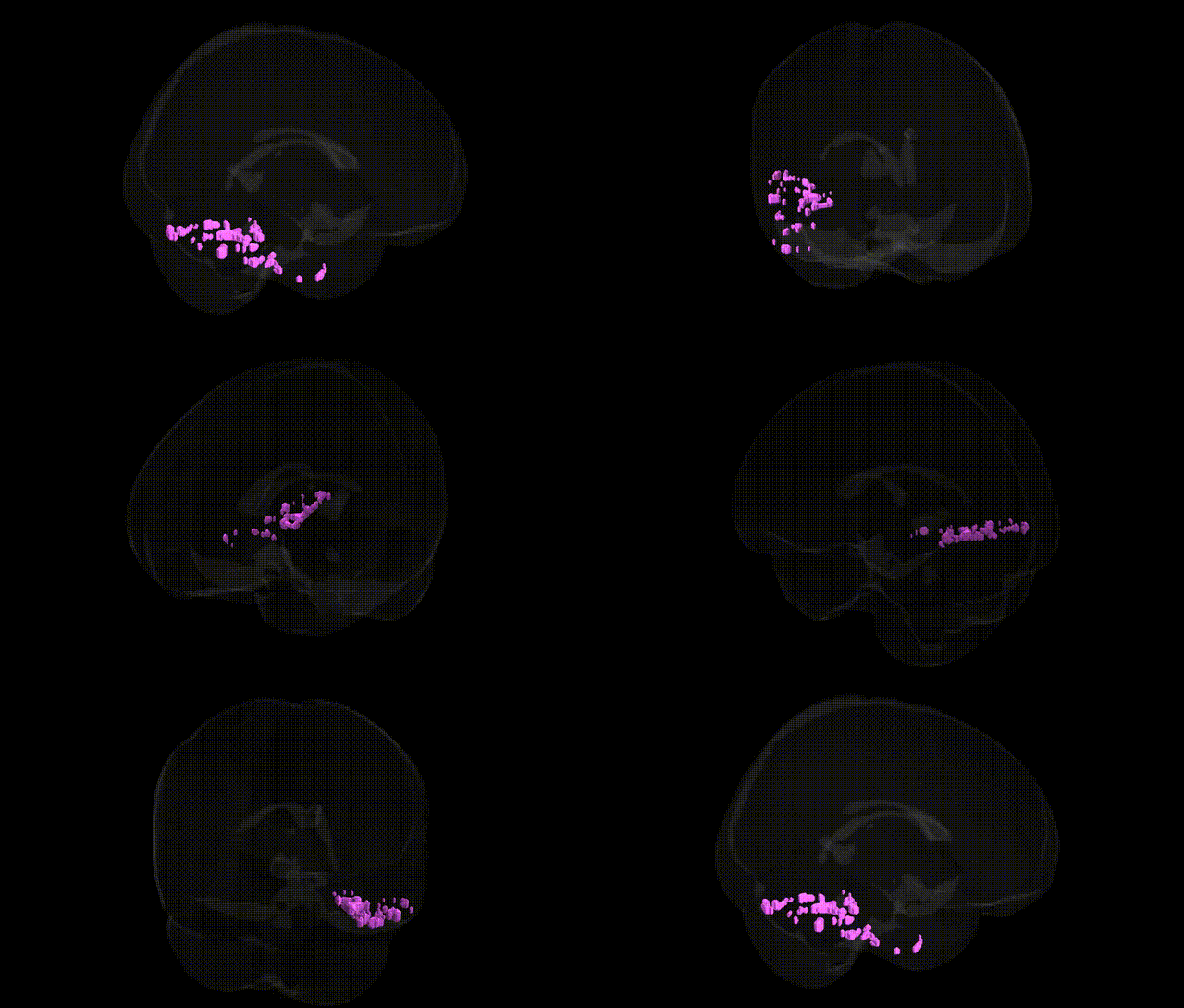
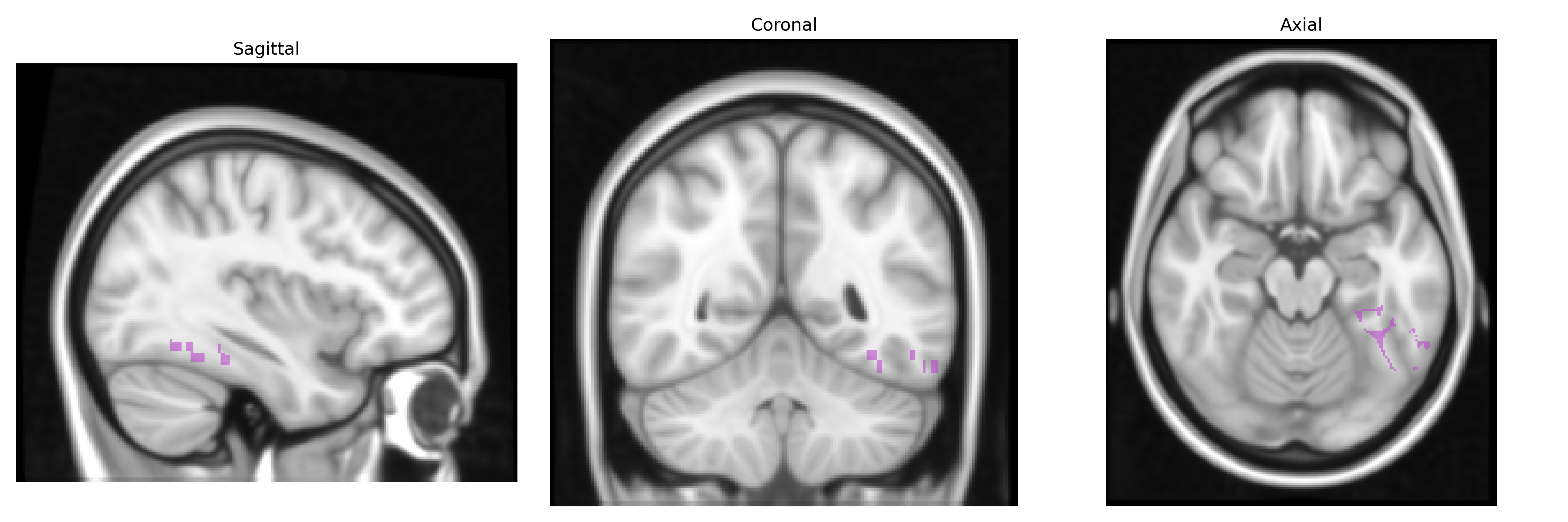
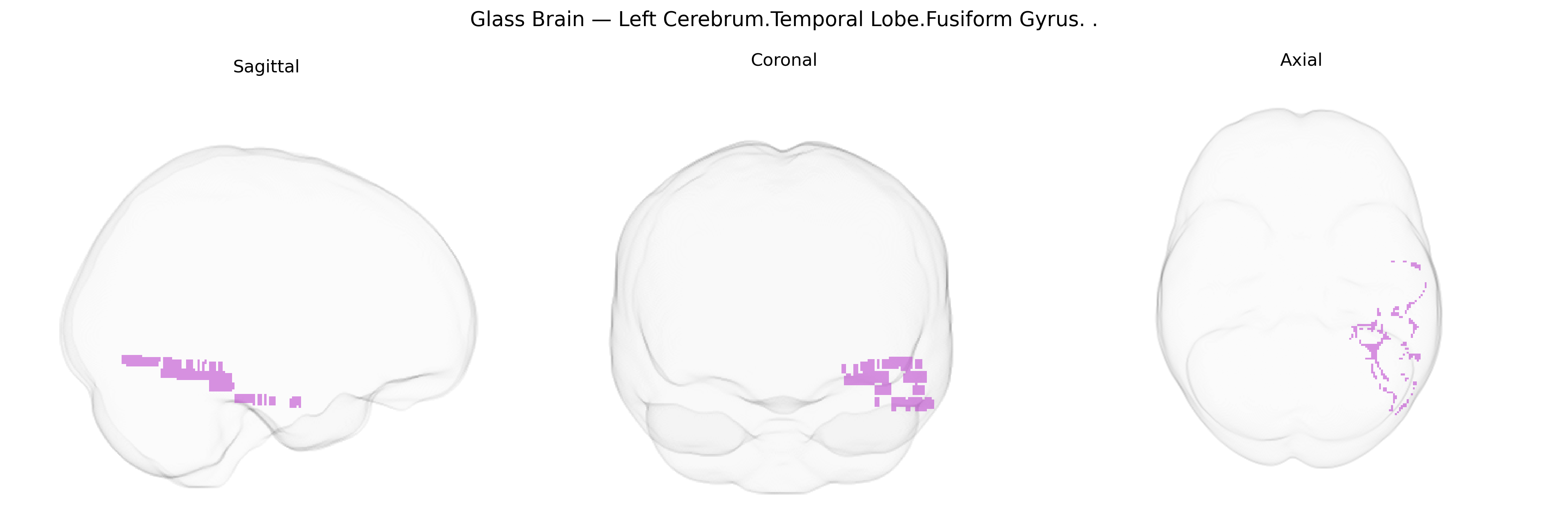
The bilateral Left Cerebrum.Temporal Lobe.Fusiform Gyrus, as defined in the Talairach 1 mm atlas, corresponds to a portion of the ventral temporal cortex located on the basal (inferior) surface of the temporal and occipital lobes. This region is cytoarchitectonically heterogeneous (e.g., encompassing areas such as FG1–FG4) and is strongly implicated in high-level visual processing, including object recognition, face perception, word and number form recognition, and complex pattern analysis. It receives extensive input from early visual areas and participates in distributed ventral stream networks supporting category-selective perception (e.g., faces, places, and written words), with lateralization of function often observed for language-related visual processing in the left hemisphere. The fusiform gyrus also shows integration with limbic and association cortices, contributing to memory-related aspects of visual recognition and semantic processing.
Fusiform gyrus
The bilateral left fusiform gyrus in the temporal lobe, as defined in the Talairach 1 mm atlas, has been repeatedly implicated in genetic and genome-wide association studies of high-level visual processing and social cognition, particularly via its role as the “fusiform face area.” Heritability studies show substantial genetic contributions to fusiform cortical thickness, surface area, and activation patterns during face and word recognition tasks, with polygenic influences overlapping those for general cognitive ability and educational attainment. GWAS consortia such as ENIGMA and UK Biobank have identified common variants in genes involved in neurodevelopment, synaptic plasticity, and axonal guidance (for example, loci near genes like RELN, BDNF, and CTNND2 in some cohorts) that associate with fusiform morphometry, although specific signals are often shared with broader temporal or ventral visual cortex measures rather than being fusiform-specific. Genetic risk for autism spectrum disorder, schizophrenia, and major depression has been linked to altered fusiform structure and function, including reduced fusiform volume or atypical face-evoked activity, with polygenic risk scores for these disorders correlating with fusiform metrics in large imaging–genetics samples. In dyslexia and other reading-related disorders, variants affecting left temporo-occipital circuits show associations with altered fusiform activation during word recognition, consistent with the region’s role as the “visual word form area,” while in prosopagnosia both rare variants and polygenic load appear to influence fusiform specialization for faces. Overall, the left fusiform gyrus emerges as a genetically influenced hub where polygenic architectures for neurodevelopmental, psychiatric, and cognitive traits converge on variation in high-level visual and social-perceptual processing.
Overview generated by GPT-4o (2026).
Region ID: 121
Hemisphere: bilateral
Atlas: Talairach labels 1mm
Full Quality Version: Download MP4
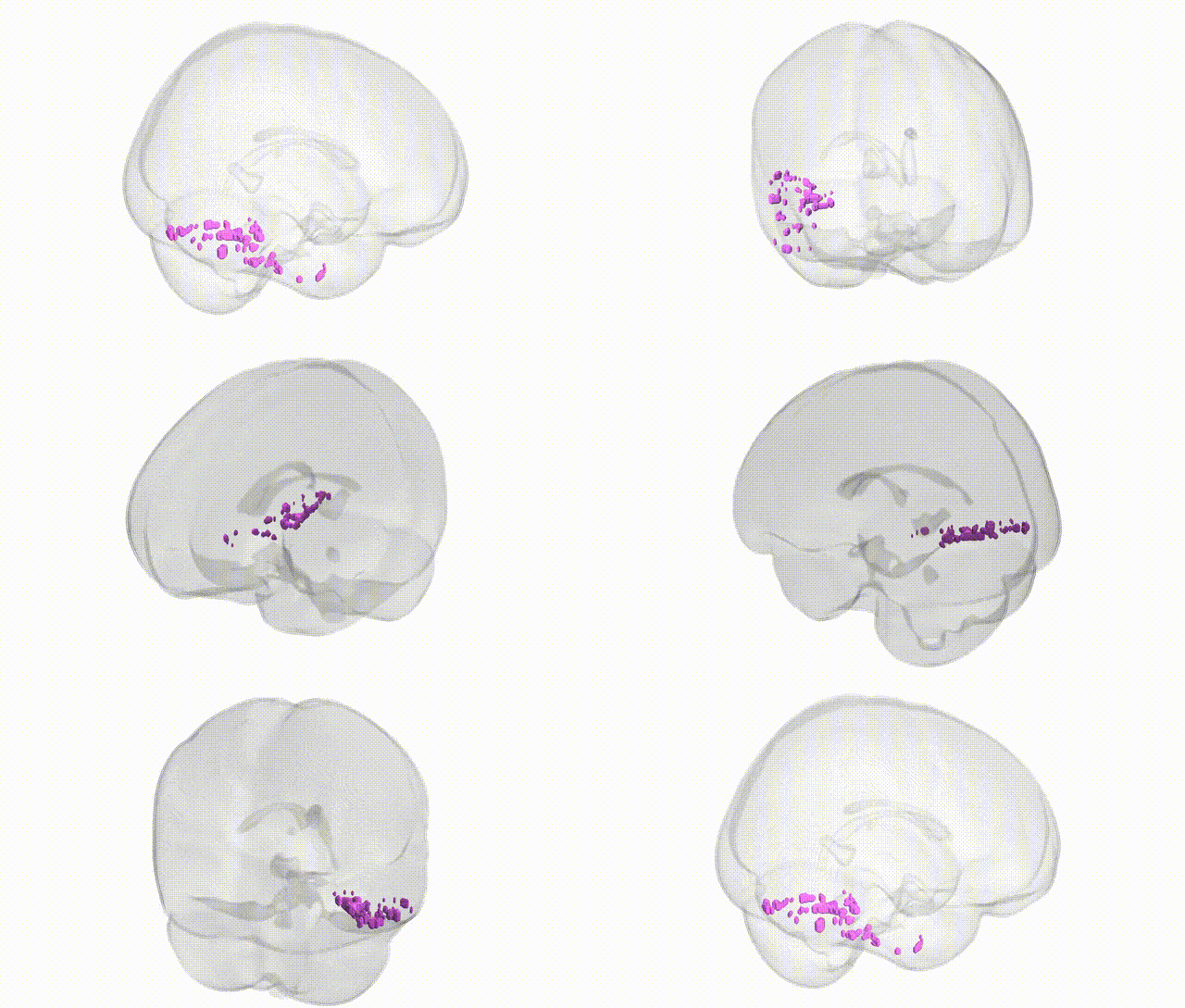
Full Quality Version: Download MP4
Wali Sidiqyar*, Gaurav Rudravaram*, Elyssa M. McMaster, Trent M. Schwartz, Adam M. Saunders, Kurt G. Schilling, Bennett A. Landman "Introducing SPINS: A Shared Public Visualization Library of Neuroanatomical Structures." Medical Imaging with Deep Learning- short paper
This resource is licensed under CC0 1.0 Universal (Public Domain).