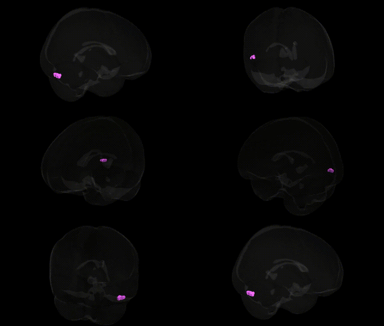
The bilateral Left Cerebrum.Temporal Lobe.Fusiform Gyrus.Gray Matter.Brodmann area 19 corresponds to a portion of the fusiform gyrus in the ventral temporal cortex that falls within the cytoarchitectonic boundaries of Brodmann area 19, part of the extrastriate visual association cortex. This region participates in higher-order visual processing, integrating complex shape, color, and pattern information and contributing to object, face, and word recognition within the ventral “what” visual pathway. Functionally, it interfaces with neighboring occipital and temporal regions, including areas involved in visual form perception and category-selective processing (such as fusiform face and word form areas), and receives inputs from primary and secondary visual cortices while projecting to downstream temporal and parietal association areas. Cytoarchitectonically, it is characterized by a well-developed granular layer IV and distinct pyramidal cell organization typical of higher-order visual association cortex. There is no direct Wikipedia article for this exact Talairach-defined region; a related structure is the Fusiform gyrus.
The bilateral left fusiform gyrus gray matter in the temporal lobe, particularly overlapping Brodmann area 19, has been implicated in genetic studies of cortical structure and neuropsychiatric traits, though most GWAS report broader occipitotemporal or fusiform measures rather than Talairach-specific labels. Large imaging-genetics consortia (e.g., ENIGMA, UK Biobank) have identified associations between fusiform or adjacent occipitotemporal cortical thickness/surface area and common variants near genes involved in neurodevelopment, synaptic function, and extracellular matrix (such as SNPs near MIR1228, WNT3, and genes in calcium channel and glutamatergic pathways), with polygenic influences overlapping general brain size and cognitive ability. Variants in genes linked to face processing and visual recognition (e.g., CDH13, NRG1, and others from GWAS of fusiform activation and cortical morphology) have been associated with functional or structural differences in this region, consistent with its role in high-level visual processing. Disorders including autism spectrum disorder, schizophrenia, major depression, and developmental dyslexia show heritable alterations of fusiform/BA19 volume or thickness, with associated risk loci often mapping to synaptic, axon guidance, or chromatin-remodeling genes (for example, GWAS hits in or near CACNA1C, ZNF804A, and multiple MHC-region genes) that also affect occipitotemporal cortices more broadly. Additionally, GWAS of visual perception, reading ability, and face recognition abilities indicate polygenic overlap with fusiform structure and activation, although no single gene has been uniquely or specifically assigned to the bilateral left fusiform BA19 region independent of neighboring visual association areas.
Overview generated by GPT-4o (2026).
Region ID: 265
Hemisphere: bilateral
Atlas: Talairach labels 1mm
Full Quality Version: Download MP4
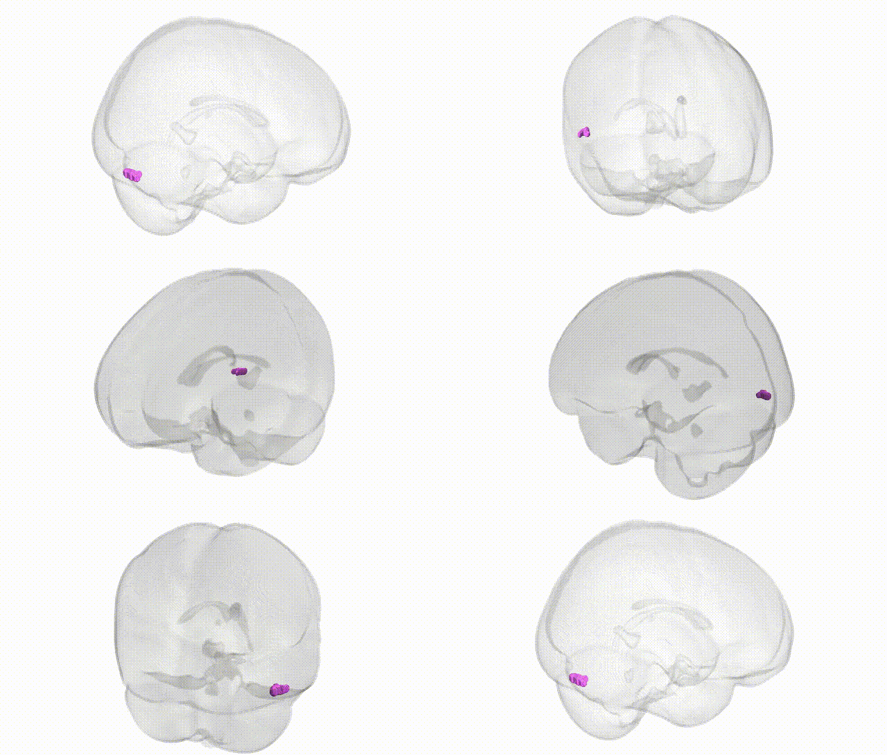
Full Quality Version: Download MP4
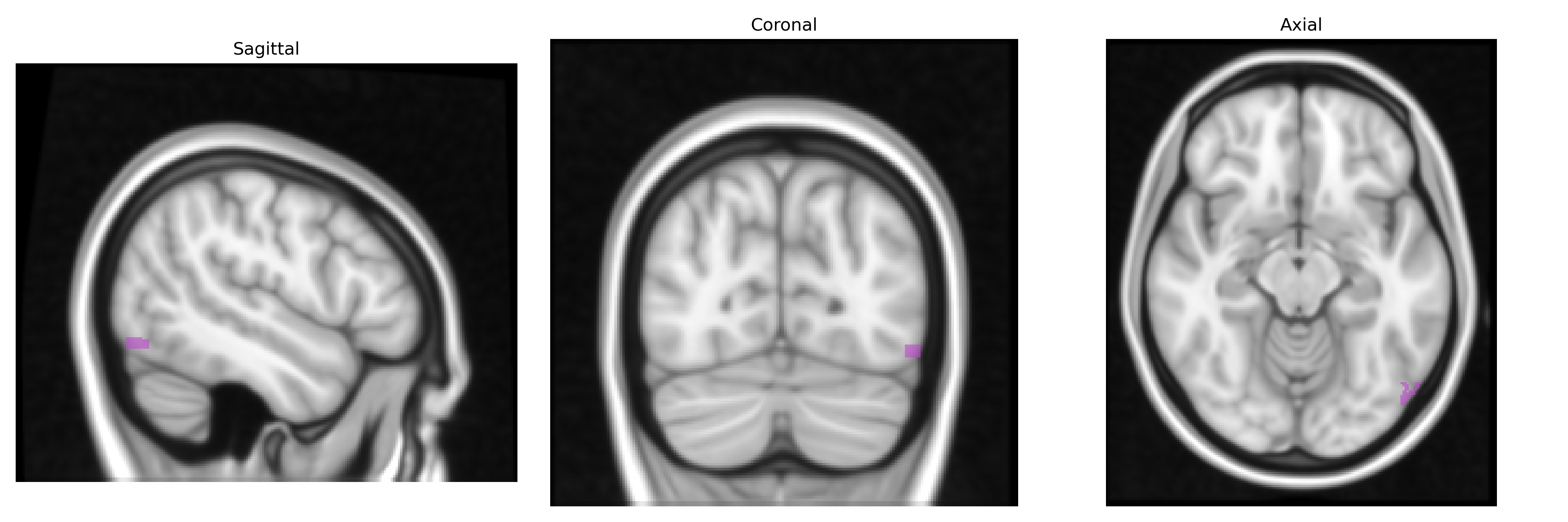
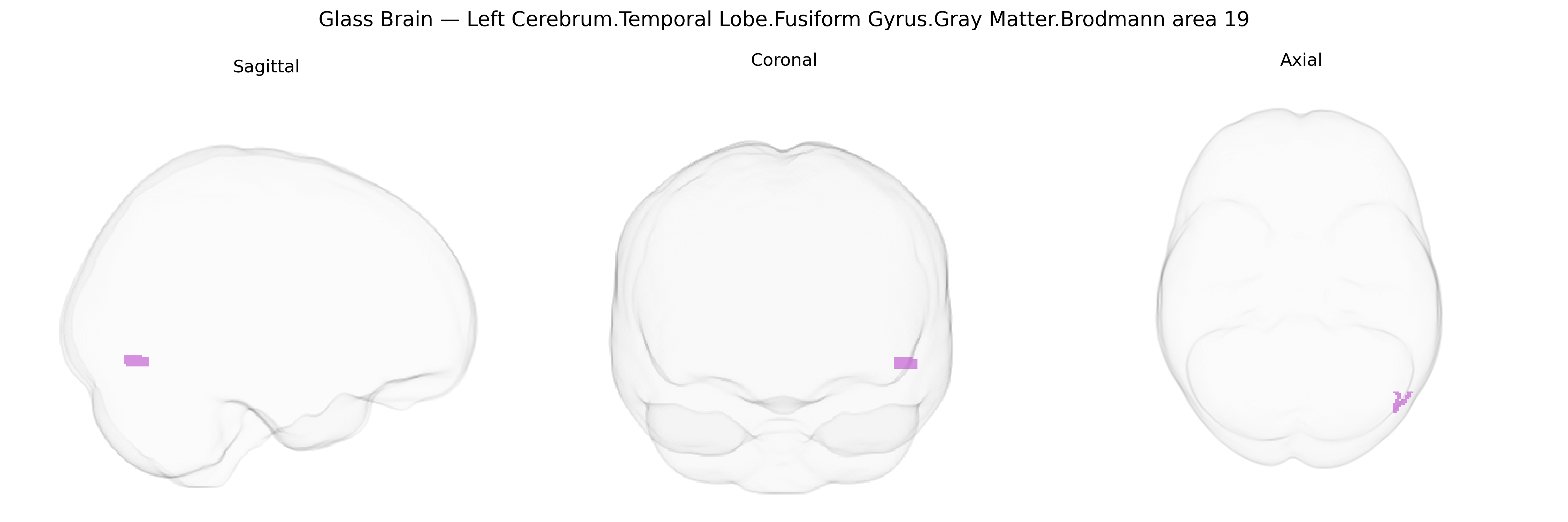
Wali Sidiqyar*, Gaurav Rudravaram*, Elyssa M. McMaster, Trent M. Schwartz, Adam M. Saunders, Kurt G. Schilling, Bennett A. Landman "Introducing SPINS: A Shared Public Visualization Library of Neuroanatomical Structures." Medical Imaging with Deep Learning- short paper
This resource is licensed under CC0 1.0 Universal (Public Domain).