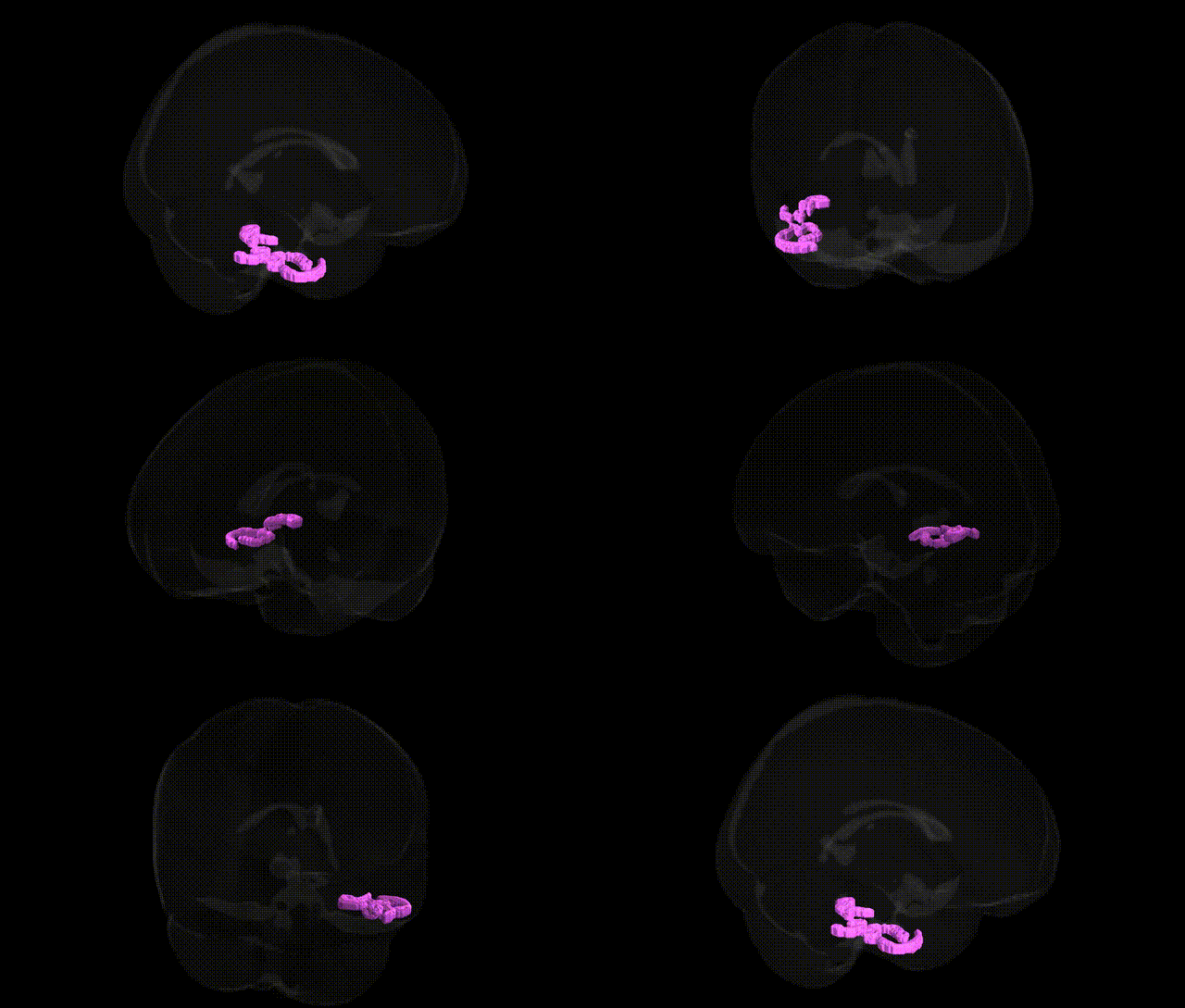
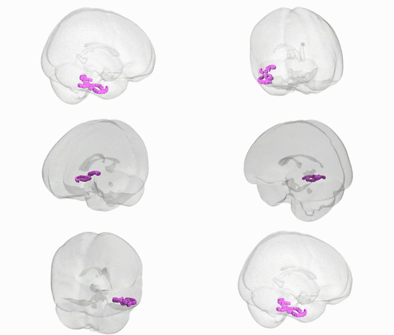
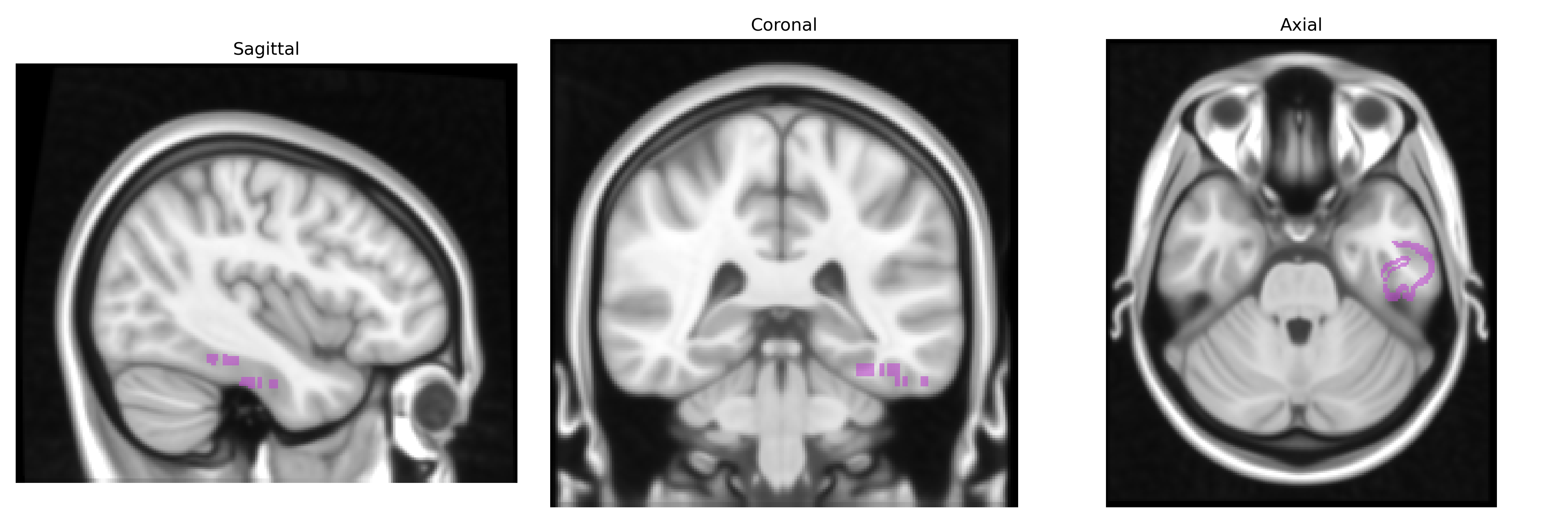
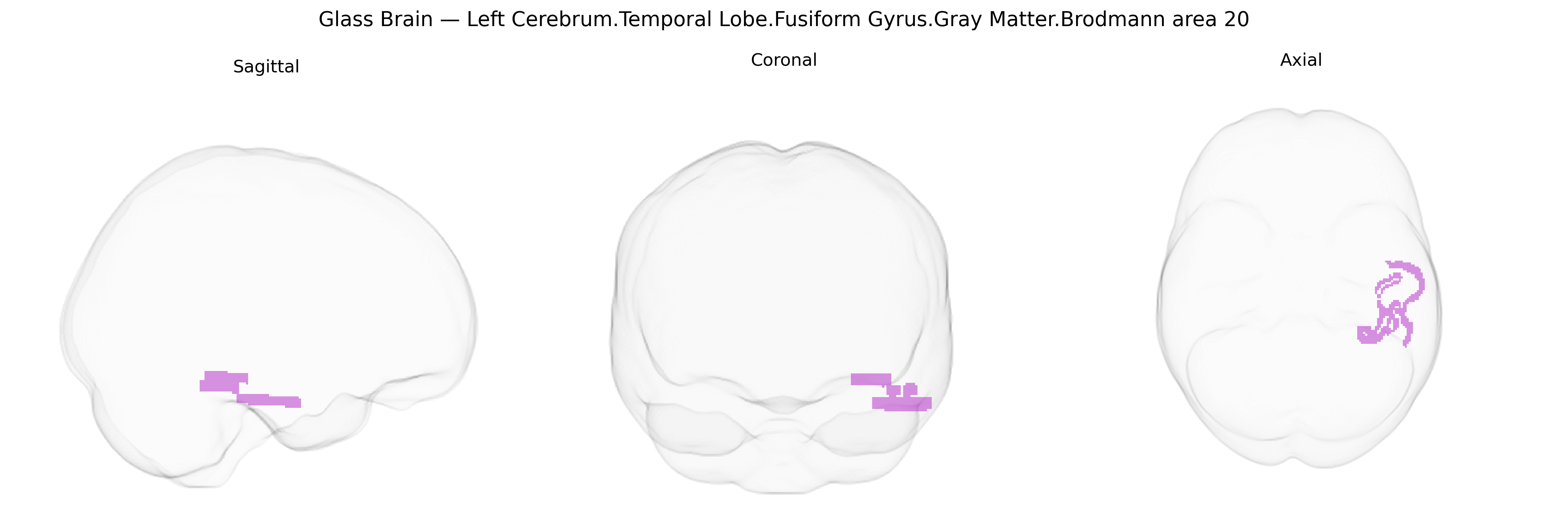
The bilateral Left Cerebrum.Temporal Lobe.Fusiform Gyrus.Gray Matter.Brodmann area 20 corresponds to a ventral temporal cortical region situated in the fusiform gyrus, typically associated with higher-order visual processing and object recognition, including the integration of complex form, color, and semantic information. As part of the inferior temporal cortex, Brodmann area 20 receives processed visual input from more posterior occipitotemporal regions and contributes to the identification and categorization of objects, faces, and words, as well as aspects of visual memory and language-related semantic processing. This region is interconnected with adjacent fusiform and inferior temporal areas, limbic structures, and frontal association cortices, forming networks important for visual perception, memory encoding and retrieval, and conceptual knowledge. There is no direct link for this exact composite label; a closely related structure is the fusiform gyrus: Fusiform gyrus.
The left fusiform gyrus (Brodmann area 20) is strongly implicated in genetic influences on higher visual and language processing, with twin and family studies indicating high heritability of fusiform volume and cortical thickness, and SNP-based studies showing significant polygenic contributions from common variants. GWAS of brain imaging phenotypes (e.g., ENIGMA, UK Biobank) have identified loci near or within genes involved in neurodevelopment and synaptic function—such as DLG2, CNTNAP2, GRIN2B, and KIAA0319—that modulate fusiform and adjacent temporal lobe gray matter structure, often overlapping with loci linked to reading ability, language skills, and general cognitive performance. Variants associated with dyslexia and specific language impairment (e.g., in DCDC2, KIAA0319, ROBO1) have been related to altered structure or activation in left temporal and fusiform regions supporting orthographic and phonological processing, while autism spectrum disorder and schizophrenia risk loci (including those in 16p11.2, 22q11.2, and genes such as NRXN1 and SHANK3) have been associated with atypical fusiform morphology or face-selective activation, consistent with social perception deficits. GWAS and candidate gene studies of prosopagnosia and face recognition ability have implicated variants affecting fusiform face area function, including loci near MCTP2 and other synaptic genes, while Alzheimer’s disease and frontotemporal dementia risk alleles (e.g., APOE ε4, MAPT haplotypes, and TREM2 variants) show associations with accelerated atrophy in left temporal and fusiform regions involved in semantic memory and object recognition. Collectively, genetic studies converge on neurodevelopmental, synaptic, and neurodegenerative pathways that influence the structure and function of the left fusiform gyrus, linking this region to language and reading traits, social cognition, major psychiatric disorders, and dementia-related neurodegeneration.
Overview generated by GPT-4o (2026).
Region ID: 123
Hemisphere: bilateral
Atlas: Talairach labels 1mm
Full Quality Version: Download MP4
Full Quality Version: Download MP4
Wali Sidiqyar*, Gaurav Rudravaram*, Elyssa M. McMaster, Trent M. Schwartz, Adam M. Saunders, Kurt G. Schilling, Bennett A. Landman "Introducing SPINS: A Shared Public Visualization Library of Neuroanatomical Structures." Medical Imaging with Deep Learning- short paper
This resource is licensed under CC0 1.0 Universal (Public Domain).