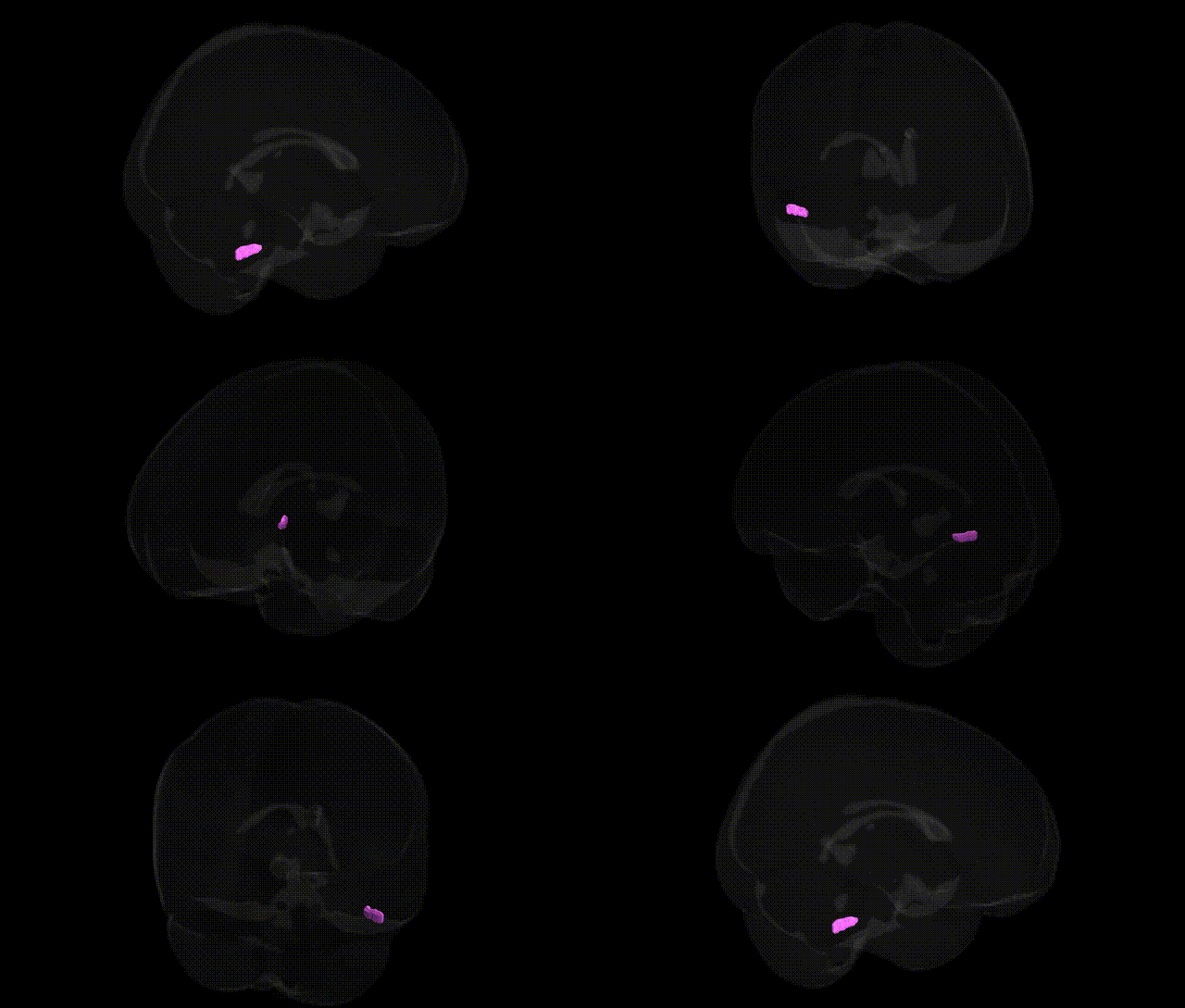
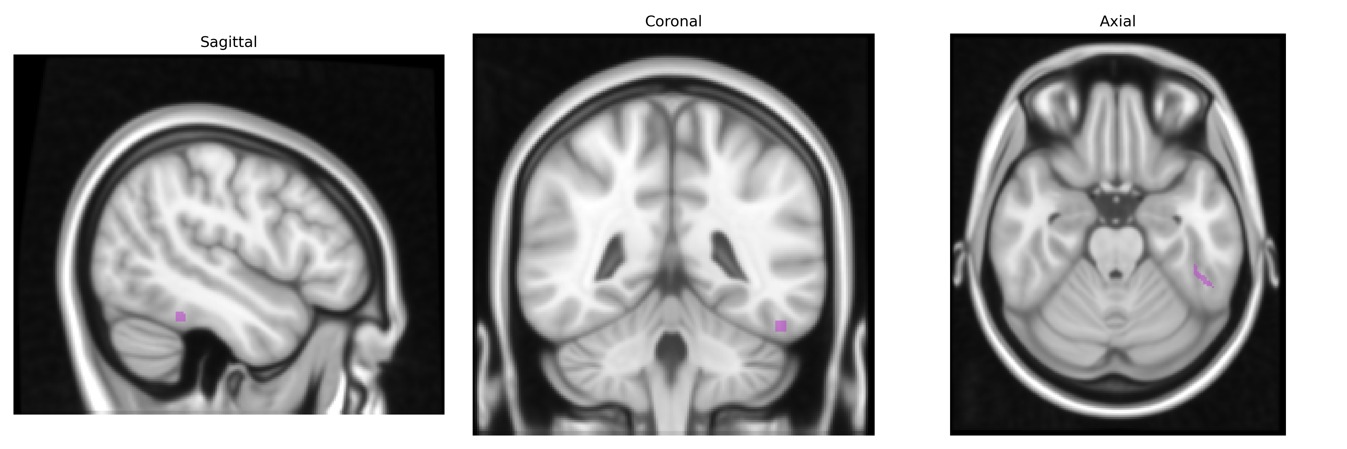
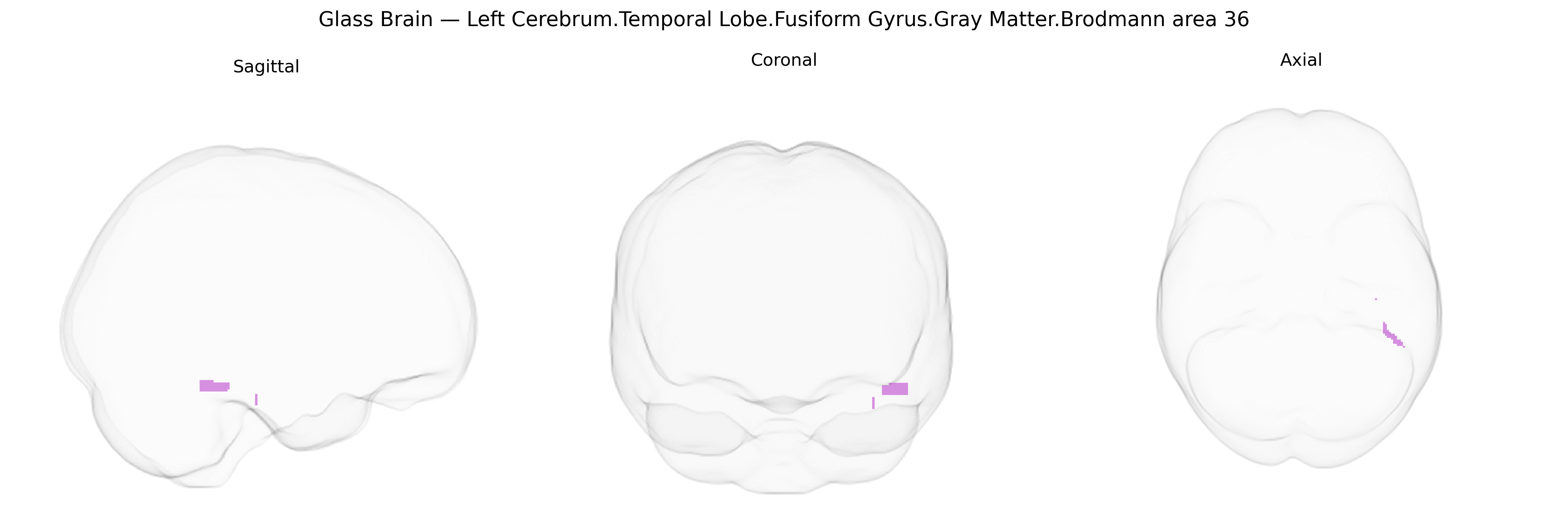
The bilateral Left Cerebrum.Temporal Lobe.Fusiform Gyrus.Gray Matter.Brodmann area 36 corresponds to a portion of the parahippocampal/lingual–fusiform region typically considered part of the ectorhinal cortex and perirhinal cortex, located on the ventromedial surface of the temporal lobe adjacent to the rhinal sulcus. Brodmann area 36 is implicated in higher-order visual processing, particularly in object recognition, complex feature conjunctions, and mnemonic functions that support familiarity-based memory and associative learning, interfacing between unimodal visual areas and medial temporal lobe memory structures. It receives input from visual association cortices and projects to hippocampal and parahippocampal regions, contributing to the encoding and retrieval of visual and multimodal representations of objects and scenes. There is no direct link for Brodmann area 36; related region: Perirhinal cortex.
The bilateral left fusiform gyrus gray matter in Brodmann area 36 (part of the temporal lobe and medial ventral visual pathway) has been implicated in several genetic and GWAS findings, largely via imaging genetics studies that link common variants to regional cortical volume, thickness, and functional activation. Large brain-structure GWAS consortia (e.g., ENIGMA, UK Biobank) have identified polygenic influences on temporal and fusiform morphology, with variants in genes such as MAPT, KIAA0586, DLG2, and multiple loci within synaptic, neurodevelopmental, and axon-guidance pathways contributing to inter-individual variation, although most associations are regionally coarse rather than Brodmann-area–specific. Fusiform and adjacent BA36 anatomy and activation have been repeatedly associated with genetic risk for neurodevelopmental and neuropsychiatric conditions, including autism spectrum disorder (with risk variants in genes such as CNTNAP2, SHANK3, and other synaptic genes tied to altered fusiform responses to faces), schizophrenia (with polygenic risk scores linked to temporal and fusiform structural differences), and dyslexia and reading-related traits (where GWAS and candidate gene studies involving DCDC2, KIAA0319, and other reading-related loci show associations with left ventral temporal language/orthographic regions that include BA36/visual word form area). In addition, Alzheimer’s disease and other dementias show genetically mediated vulnerability in medial temporal structures incorporating BA36, with APOE ε4 and other AD risk loci associated with accelerated atrophy and connectivity changes in perirhinal/entorhinal-fusiform circuits. Overall, genetic architecture affecting synaptic function, cortical development, and neurodegeneration appears to modulate the structure and function of the BA36 fusiform region, contributing to risk and phenotypic variation in social cognition, language/reading, memory, and higher-order visual processing.
Overview generated by GPT-4o (2026).
Region ID: 136
Hemisphere: bilateral
Atlas: Talairach labels 1mm
Full Quality Version: Download MP4
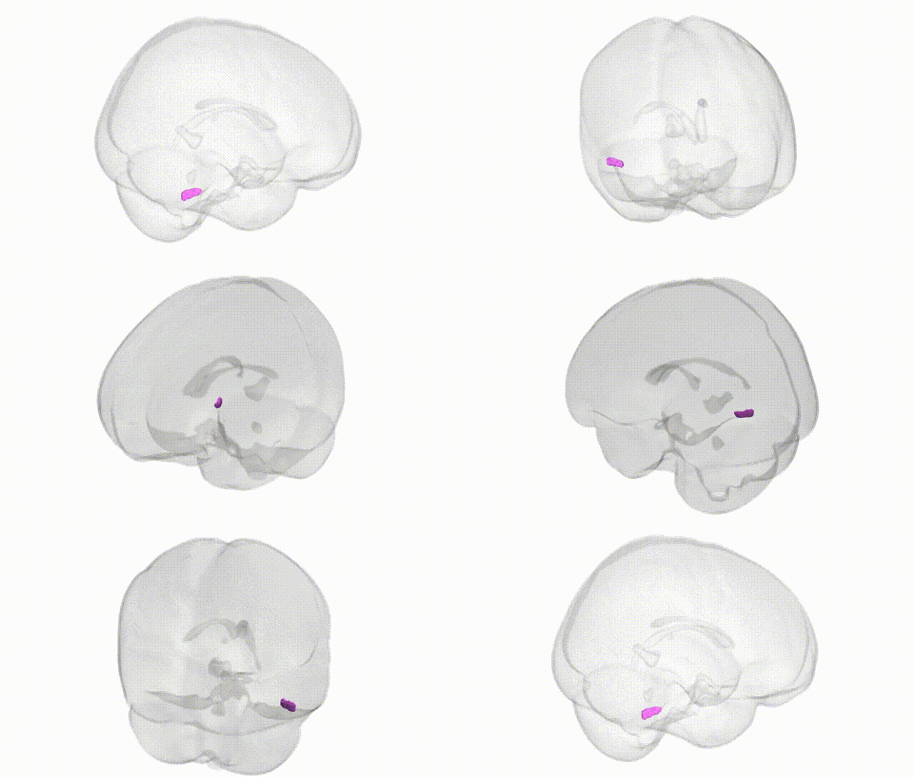
Full Quality Version: Download MP4
Wali Sidiqyar*, Gaurav Rudravaram*, Elyssa M. McMaster, Trent M. Schwartz, Adam M. Saunders, Kurt G. Schilling, Bennett A. Landman "Introducing SPINS: A Shared Public Visualization Library of Neuroanatomical Structures." Medical Imaging with Deep Learning- short paper
This resource is licensed under CC0 1.0 Universal (Public Domain).