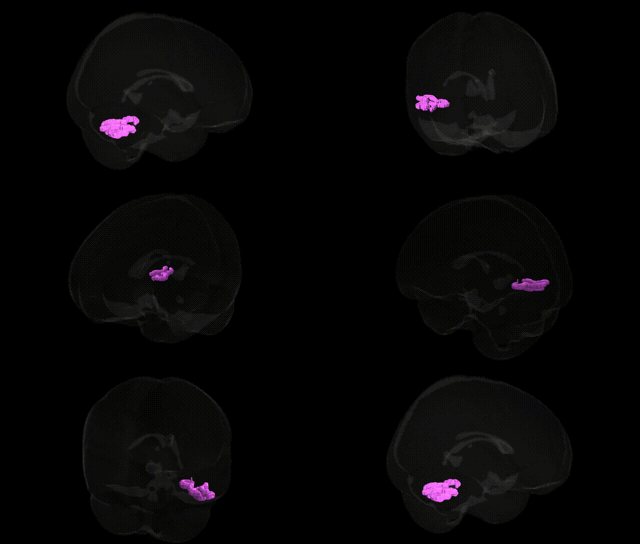
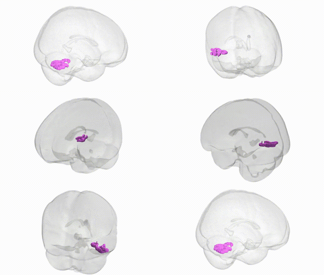
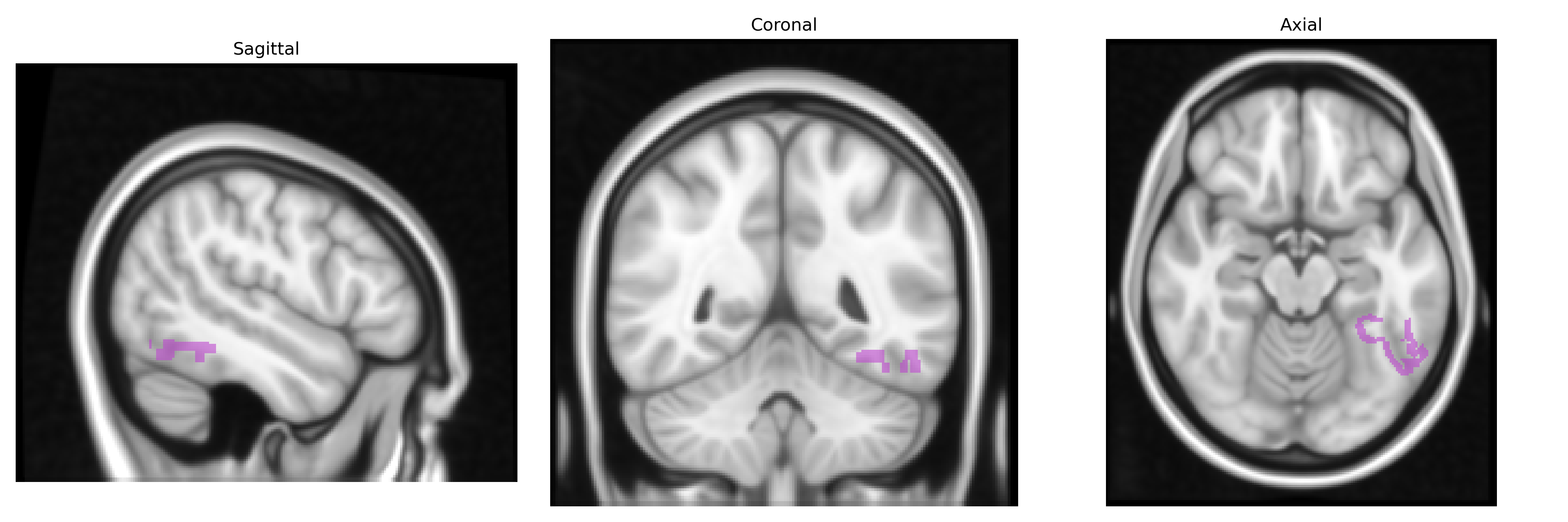
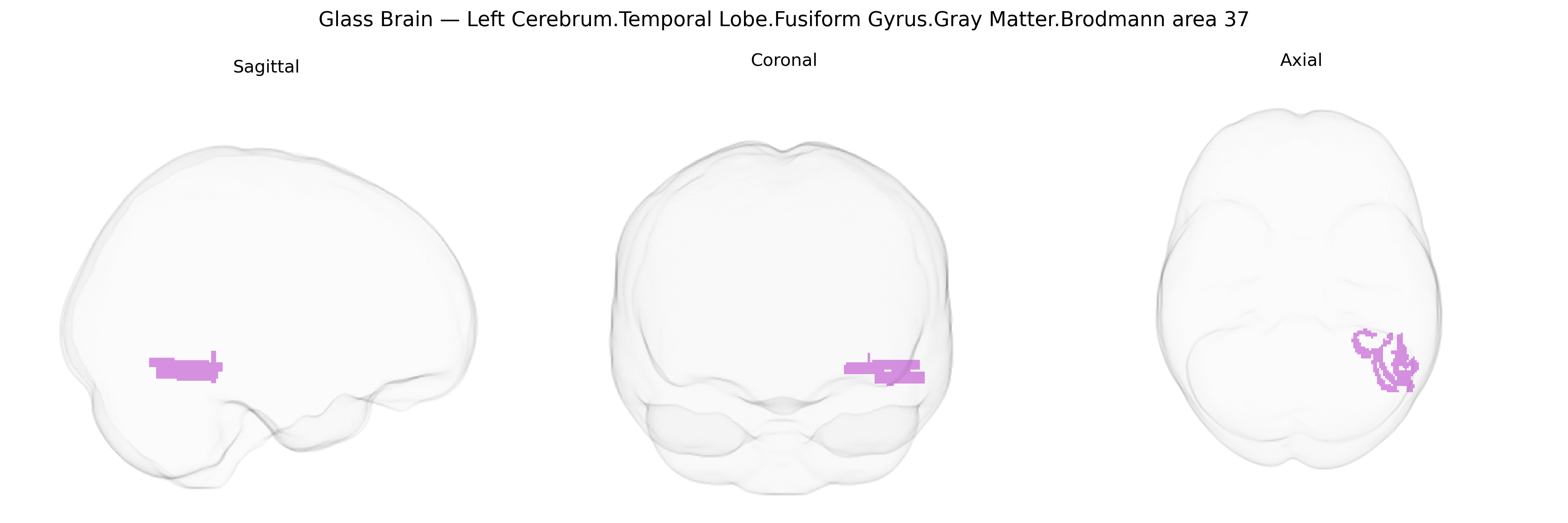
The bilateral Left Cerebrum.Temporal Lobe.Fusiform Gyrus.Gray Matter.Brodmann area 37 corresponds to a ventral temporal cortical region in the fusiform gyrus, classically associated with high-level visual processing and category-specific recognition. Brodmann area 37 lies posterolaterally in the temporal lobe, bordering occipitotemporal regions, and is strongly implicated in object, word, and especially face processing, contributing to visual memory, semantic integration, and the linkage of visual form with linguistic and conceptual representations. Functionally, this region participates in the ventral “what” pathway, integrating detailed visual features into coherent percepts and supporting tasks such as reading, recognition of familiar objects and faces, and retrieval of associated semantic knowledge; lesions can lead to deficits like visual agnosia or category-specific recognition impairments. Fusiform gyrus
The bilateral left fusiform gyrus in Brodmann area 37, as defined in the Talairach 1 mm atlas, has been implicated in genetic studies of both structure and function, particularly in relation to fusiform volume, cortical thickness, and specialized roles in face and word processing. Twin and SNP-based heritability studies indicate substantial genetic influence on fusiform gray matter volume and surface area, with genome-wide association analyses of brain imaging traits (e.g., ENIGMA, UK Biobank) identifying common variants in genes involved in neurodevelopment, synaptic function, and cortical patterning that affect temporal and ventral occipitotemporal cortex, including regions overlapping BA37. Genetic associations involving this region have been reported for neurodevelopmental and psychiatric conditions such as autism spectrum disorder (where altered fusiform structure and hypoactivation during face processing co-occur with risk variants in synaptic and neurodevelopmental genes), schizophrenia (where temporal and fusiform gray matter reductions and altered activation correlate with polygenic risk), and dyslexia or reading-related traits (where BA37/ventral occipitotemporal cortex, including the visual word form area, shows structural and functional changes linked to variants in genes such as DCDC2 and KIAA0319 in broader reading networks). Additional GWAS and imaging-genetics work ties fusiform/BA37 morphology and activation to face recognition ability, social cognition, and aspects of language and semantic processing, suggesting that polygenic variation shaping ventral temporal cortex development and connectivity contributes to individual differences and disorder risk in domains subserved by this region.
Overview generated by GPT-4o (2026).
Region ID: 208
Hemisphere: bilateral
Atlas: Talairach labels 1mm
Full Quality Version: Download MP4
Full Quality Version: Download MP4
Wali Sidiqyar*, Gaurav Rudravaram*, Elyssa M. McMaster, Trent M. Schwartz, Adam M. Saunders, Kurt G. Schilling, Bennett A. Landman "Introducing SPINS: A Shared Public Visualization Library of Neuroanatomical Structures." Medical Imaging with Deep Learning- short paper
This resource is licensed under CC0 1.0 Universal (Public Domain).