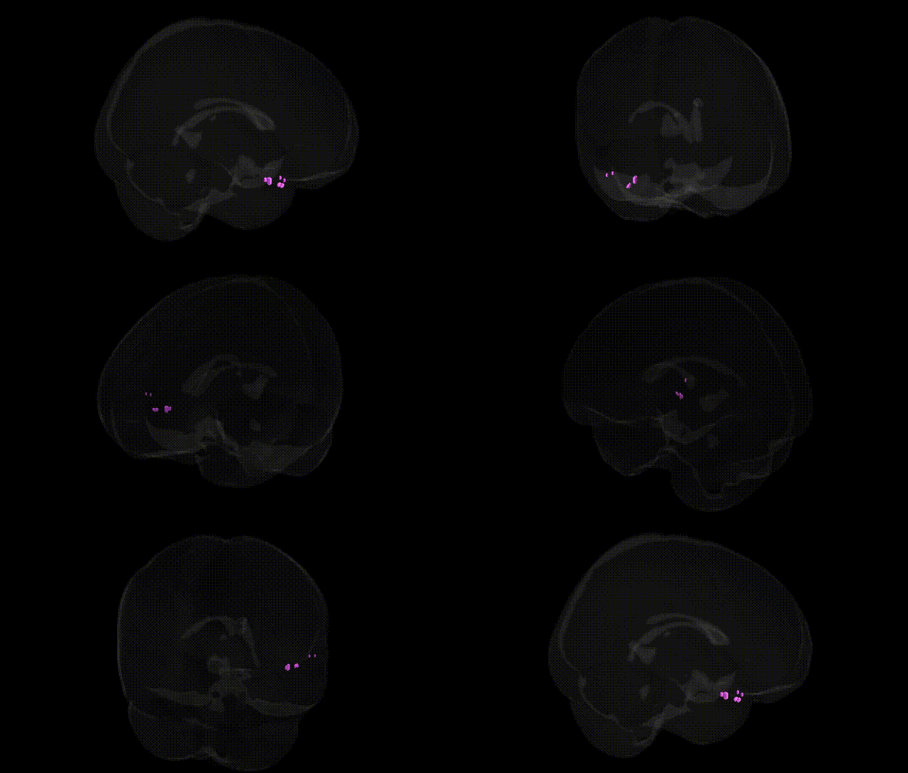
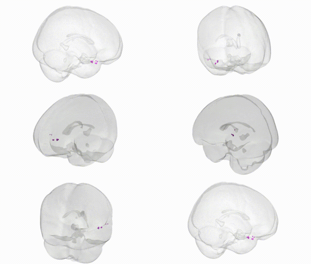
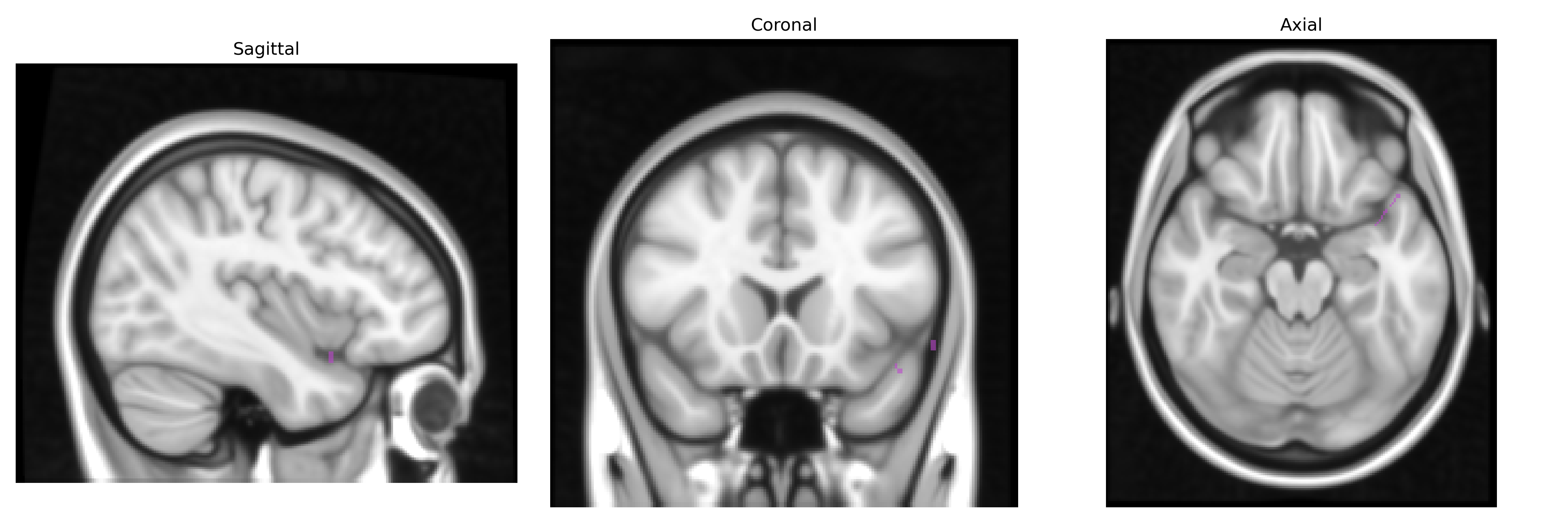
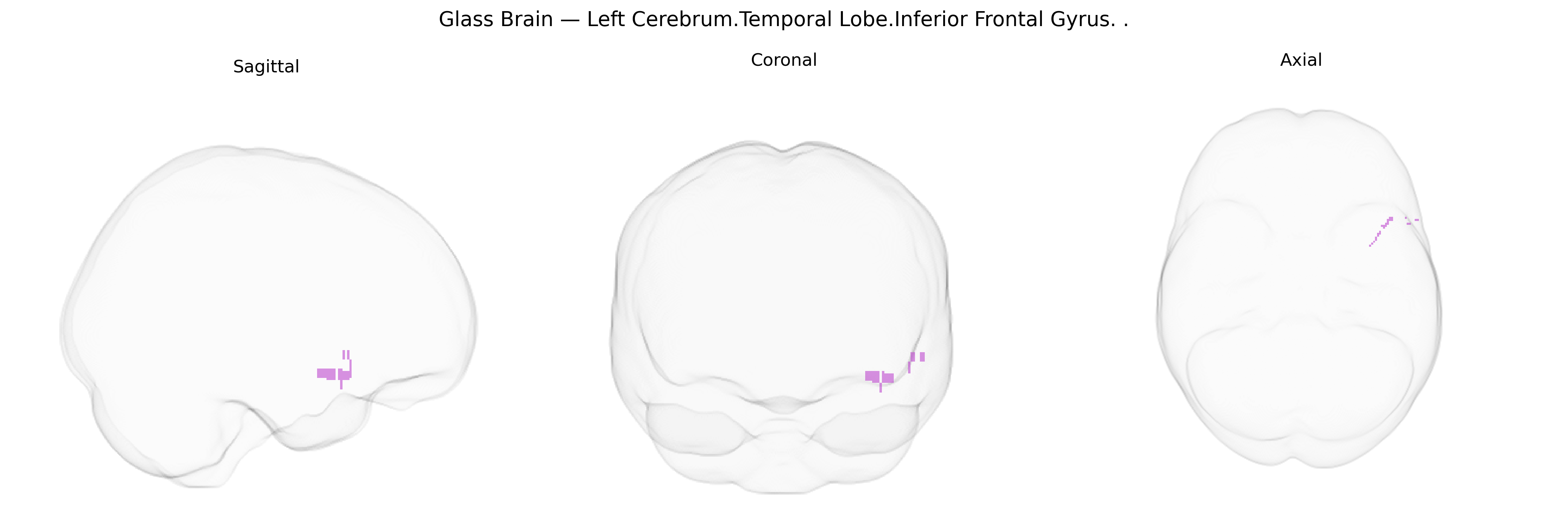
The bilateral Left Cerebrum.Temporal Lobe.Inferior Frontal Gyrus region, as referenced in the Talairach 1 mm atlas, corresponds primarily to portions of the inferior frontal gyrus (notably encompassing parts of the pars opercularis and pars triangularis) that border or interface with anterior temporal regions through fronto-temporal association pathways. Functionally, this area is heavily involved in language production and processing (classically overlapping with Broca’s area in the dominant hemisphere), semantic retrieval, syntactic processing, and aspects of controlled speech and verbal working memory, while also contributing to higher-order executive functions such as response inhibition and cognitive control. Through extensive connections with temporal lobe structures (including superior and middle temporal gyri), this region participates in integrating auditory-linguistic input with articulatory and executive processes, supporting coherent speech and complex language comprehension.
The bilateral left inferior frontal gyrus (IFG) within the temporal/frontal lobe—corresponding to language-related areas such as pars opercularis/triangularis in the Talairach 1 mm atlas—has been repeatedly implicated in genetic studies of language, cognition, and psychiatric/neurodevelopmental disorders. Twin and GWAS-based heritability work on cortical thickness, surface area, and sulcal morphology shows significant genetic influences on IFG structure, with several large consortia (e.g., ENIGMA) identifying loci that affect left IFG volume and asymmetry, often in genes involved in neuronal migration, axon guidance, synaptic plasticity, and transcriptional regulation. FOXP2, CNTNAP2, and related language-network genes have been associated with functional activation and connectivity changes in this region, particularly in tasks involving speech production, phonological processing, and syntactic computation. Polygenic risk for schizophrenia, bipolar disorder, and major depressive disorder has been linked to altered IFG structure and activation, and imaging–genetics studies show that risk variants in genes such as ZNF804A and DISC1 modulate IFG activity during working memory and language tasks. Autistic traits and ADHD have also been tied to genetic variation influencing inferior frontal anatomy and functional coupling with temporal and parietal language networks. Additionally, GWAS of reading ability, dyslexia, and educational attainment implicate loci whose effects partly converge on left IFG morphology and activation, underscoring this region’s genetically influenced role in language, executive control, and higher cognition.
Overview generated by GPT-4o (2026).
Region ID: 232
Hemisphere: bilateral
Atlas: Talairach labels 1mm
Full Quality Version: Download MP4
Full Quality Version: Download MP4
Wali Sidiqyar*, Gaurav Rudravaram*, Elyssa M. McMaster, Trent M. Schwartz, Adam M. Saunders, Kurt G. Schilling, Bennett A. Landman "Introducing SPINS: A Shared Public Visualization Library of Neuroanatomical Structures." Medical Imaging with Deep Learning- short paper
This resource is licensed under CC0 1.0 Universal (Public Domain).