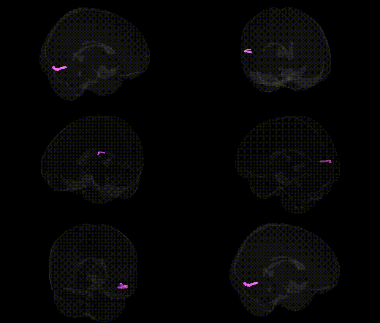
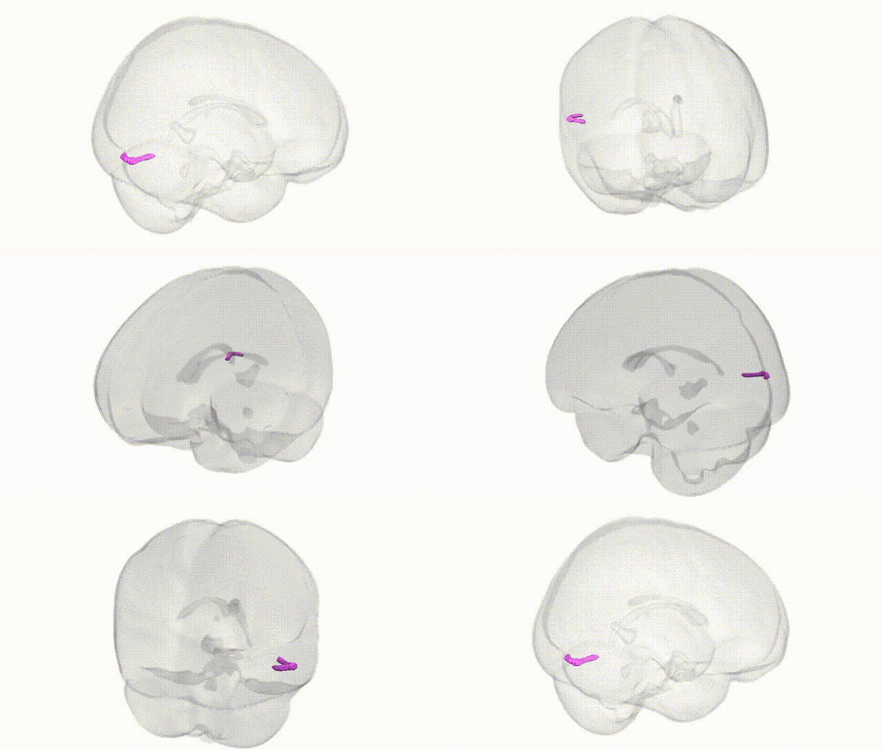
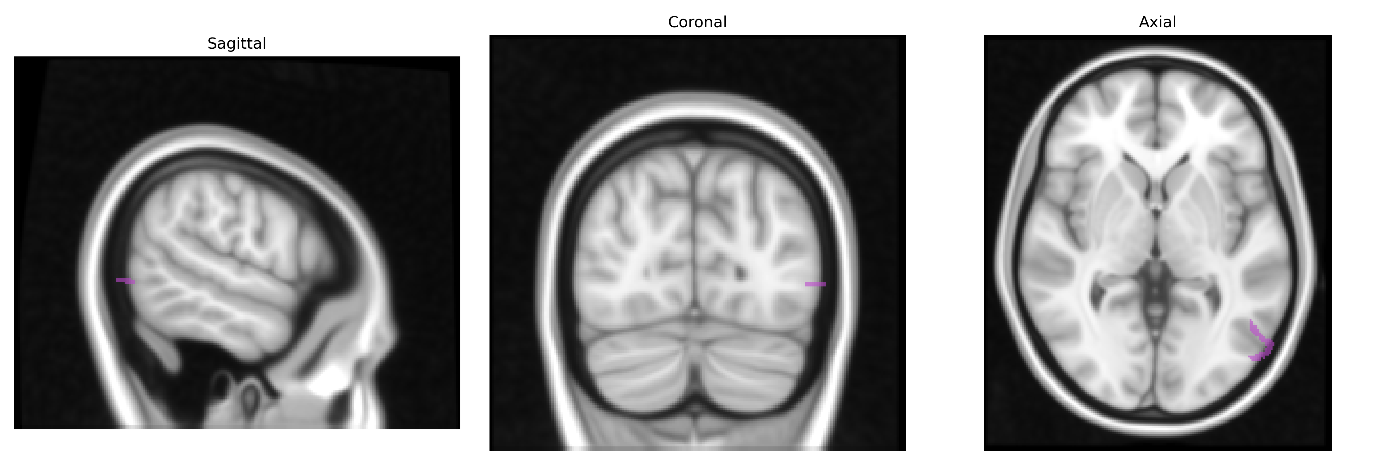
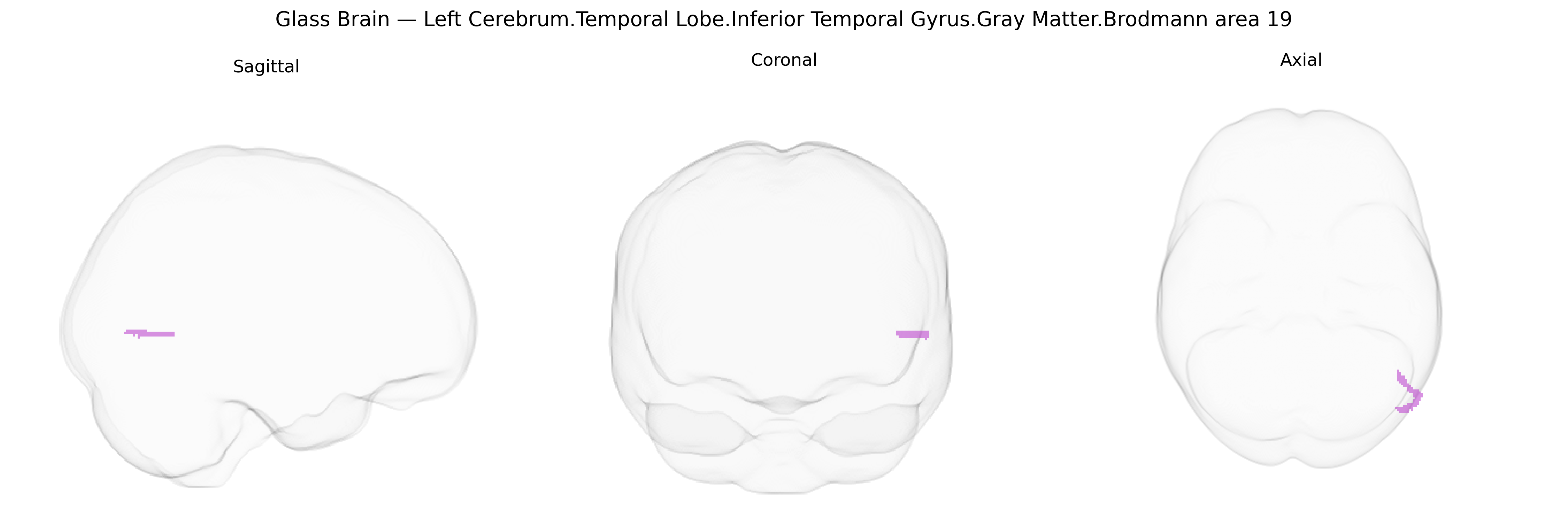
The bilateral Left Cerebrum.Temporal Lobe.Inferior Temporal Gyrus.Gray Matter.Brodmann area 19 corresponds to associative visual cortex located in the posterior portion of the inferior temporal gyrus, functionally part of the ventral visual processing stream. As a subdivision of Brodmann area 19, it participates in higher-order integration of visual information, including complex form, object, and potentially category-related processing, through extensive reciprocal connections with primary and secondary visual areas, adjacent temporal association cortices, and multimodal parietal regions. Neuronal populations in this region exhibit relatively large, complex receptive fields and contribute to transforming retinotopic visual input into more abstract, invariant representations, supporting object recognition and visually guided behavior. There is no direct link for this specific subregion; a related structure is Brodmann area 19.
The bilateral left cerebrum temporal lobe inferior temporal gyrus gray matter corresponding to Brodmann area 19 (Talairach 1 mm atlas) lies at the occipito-temporal junction and has been implicated in genetic studies primarily through imaging-genetics and GWAS of cortical thickness, surface area, and volume in occipital–temporal visual association cortex. Large consortia such as ENIGMA and UK Biobank have reported common variants in genes involved in neurodevelopment, synaptic function, and neuronal differentiation (for example, loci near HMGA2, MIR924HG, ZIC family genes, and other regulatory elements) associated with structural variation in nearby occipital/ventral temporal regions, though effects are typically small and spatially coarse rather than specific to this exact label. Functionally, this region overlaps ventral visual and higher-order association areas, and genetic risk variants for neurodevelopmental and psychiatric conditions—particularly autism spectrum disorder, schizophrenia, major depression, and dyslexia—have been linked in imaging-genetic work to altered thickness or activation in inferior and middle temporal gyri and adjacent BA19/BA37 territory, including genes affecting synaptic plasticity and axon guidance (e.g., CNTNAP2, NRG1/ERBB signaling components, and several polygenic risk scores for schizophrenia and ASD). In neurodegenerative disease, APOE ε4 and other Alzheimer’s disease risk loci (such as CLU, PICALM, BIN1) are associated with atrophy patterns that include posterior temporal–occipital association cortex overlapping BA19. Overall, current evidence ties this region to polygenic influences on visual association and higher-order cognitive networks, but no single gene or variant is uniquely or selectively associated with the specific bilateral “Left Cerebrum.Temporal Lobe.Inferior Temporal Gyrus.Gray Matter.BA19” parcel beyond these broader, distributed genetic effects.
Overview generated by GPT-4o (2026).
Region ID: 479
Hemisphere: bilateral
Atlas: Talairach labels 1mm
Full Quality Version: Download MP4
Full Quality Version: Download MP4
Wali Sidiqyar*, Gaurav Rudravaram*, Elyssa M. McMaster, Trent M. Schwartz, Adam M. Saunders, Kurt G. Schilling, Bennett A. Landman "Introducing SPINS: A Shared Public Visualization Library of Neuroanatomical Structures." Medical Imaging with Deep Learning- short paper
This resource is licensed under CC0 1.0 Universal (Public Domain).