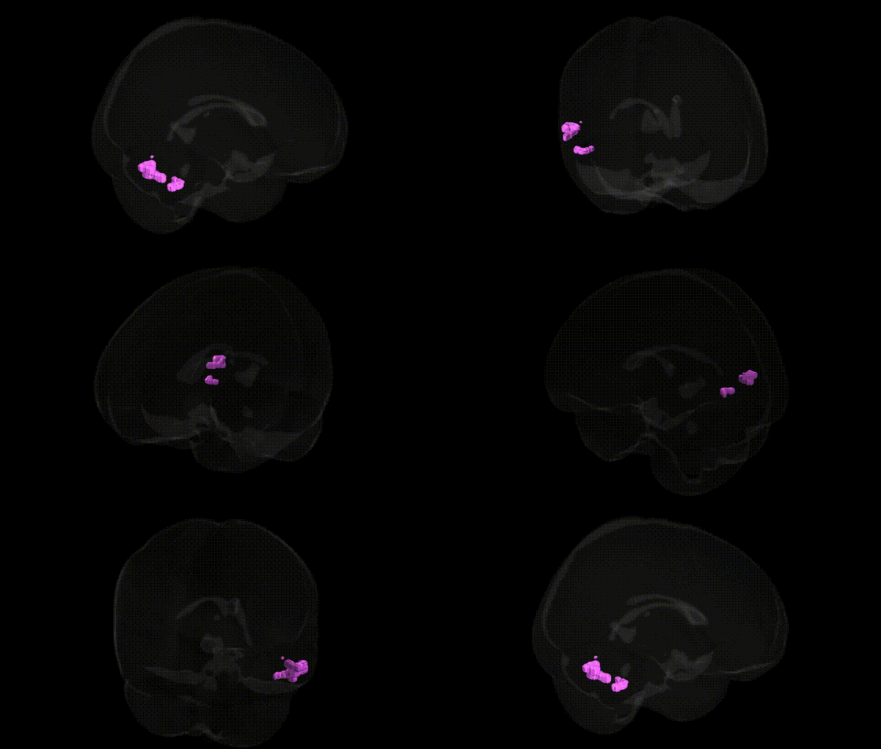
The bilateral Left Cerebrum.Temporal Lobe.Inferior Temporal Gyrus.Gray Matter.Brodmann area 37 corresponds primarily to the occipitotemporal (fusiform and adjacent inferior temporal) cortex, a high‐order visual association region implicated in object, face, and word-form recognition. Histologically defined by Brodmann’s cytoarchitectonic criteria, area 37 lies at the junction of the temporal and occipital lobes and receives convergent input from earlier visual areas, supporting complex feature integration and category-specific processing (e.g., faces, written words, and other complex visual stimuli). Functionally, this region is critical for visual semantic processing and has been associated with the ventral “what” visual pathway, contributing to the identification and recognition of objects and their meaning, with lesions often producing category-specific agnosias and impairments in reading or face recognition. There is no direct link for this exact composite region; see the related area Brodmann area 37.
The bilateral left cerebrum temporal lobe inferior temporal gyrus gray matter Brodmann area 37 (BA37), corresponding largely to the fusiform and adjacent inferior temporal cortex, has been implicated in genetic studies of face and object recognition, language-related processing, and neurodevelopmental and neurodegenerative disorders. Twin and SNP-based heritability analyses from large imaging-genetics consortia (for example, ENIGMA and UK Biobank) show moderate to high heritability of cortical thickness and surface area in this region, with genome-wide significant associations involving genes related to neuronal development, synaptic plasticity, and axon guidance (such as variants near MAPT, WNT and semaphorin pathway genes, and loci overlapping general intracranial and temporal lobe volume hits). GWAS of autism spectrum disorder, dyslexia, and language impairment have linked risk loci to altered structure or activation in BA37, particularly in left-lateralized temporal and fusiform regions supporting visual word and face processing. In schizophrenia and bipolar disorder, polygenic risk scores correlate with reduced gray matter volume or cortical thickness in inferior temporal and fusiform areas, including BA37, while Alzheimer’s disease and frontotemporal dementia genetics (for example, APOE and MAPT haplotypes) have been associated with atrophy patterns that extend into this territory. Imaging-genetics work on face recognition ability (including studies of prosopagnosia) and reading-related traits (such as variation in the visual word form area, located within BA37) suggests that common variants in genes affecting cortical patterning and myelination contribute to inter-individual differences in structure and function of this region, although specific loci with robust, replicated, region-exclusive effects remain limited and most findings reflect shared polygenic influences on broader temporal and occipitotemporal networks rather than BA37 alone.
Overview generated by GPT-4o (2026).
Region ID: 213
Hemisphere: bilateral
Atlas: Talairach labels 1mm
Full Quality Version: Download MP4
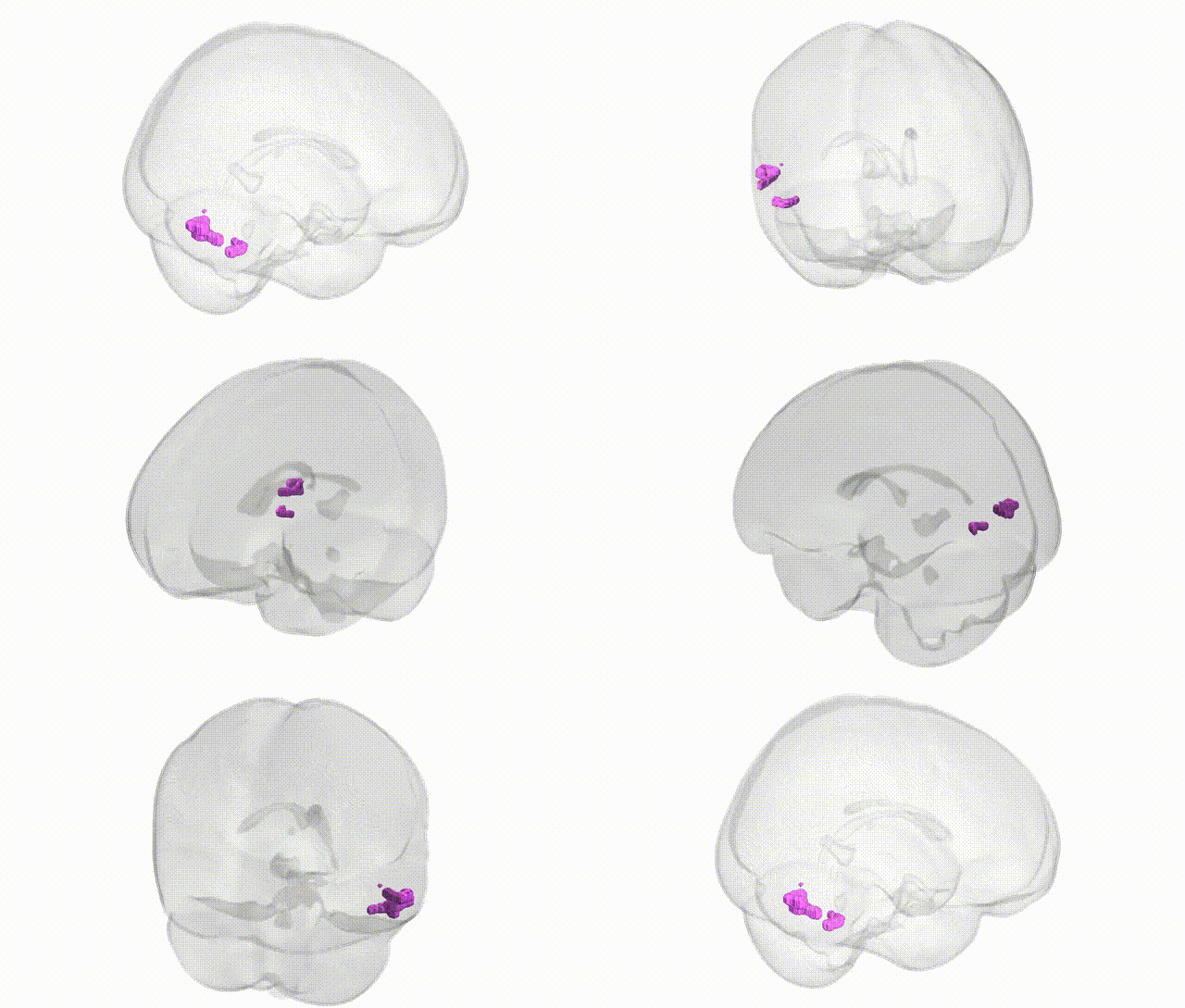
Full Quality Version: Download MP4
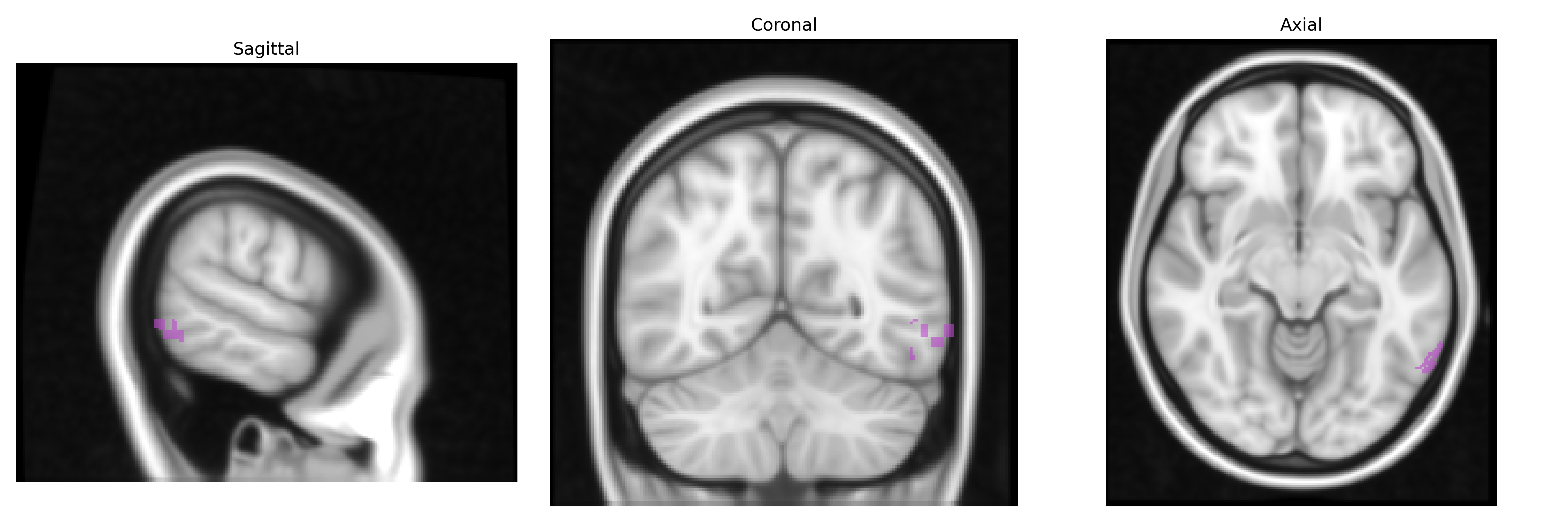
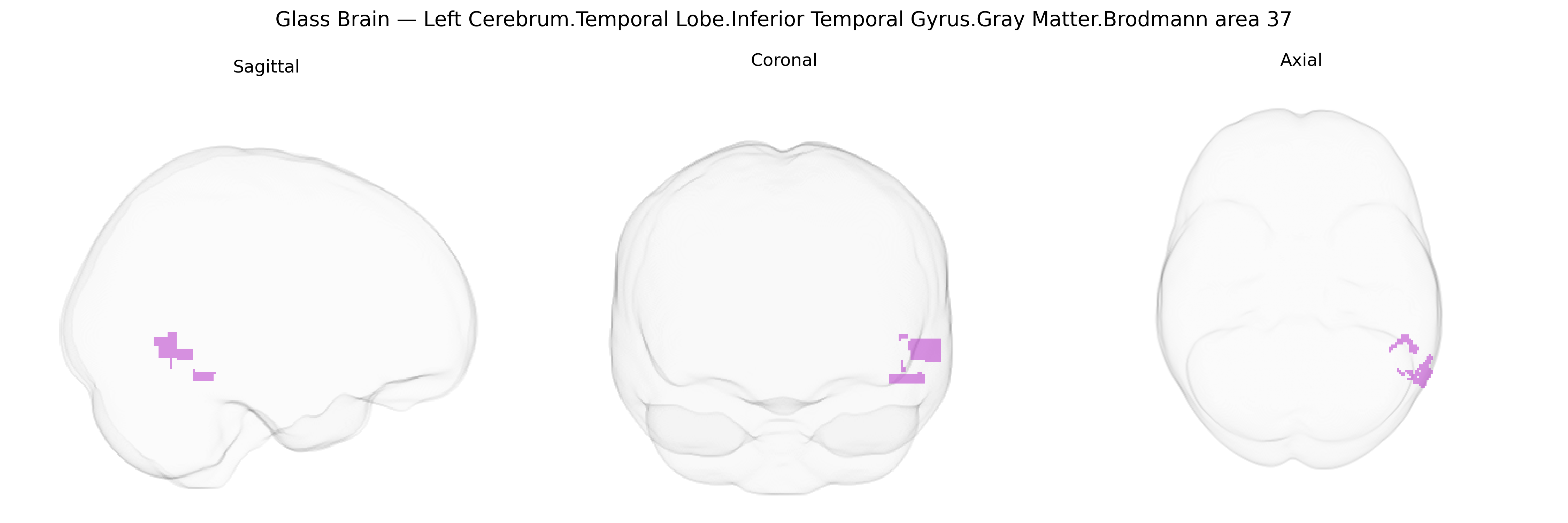
Wali Sidiqyar*, Gaurav Rudravaram*, Elyssa M. McMaster, Trent M. Schwartz, Adam M. Saunders, Kurt G. Schilling, Bennett A. Landman "Introducing SPINS: A Shared Public Visualization Library of Neuroanatomical Structures." Medical Imaging with Deep Learning- short paper
This resource is licensed under CC0 1.0 Universal (Public Domain).