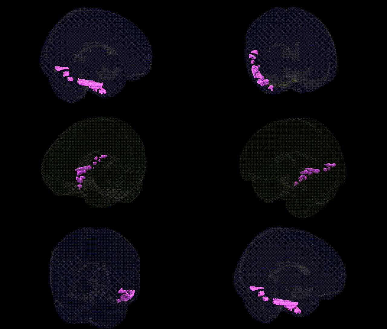
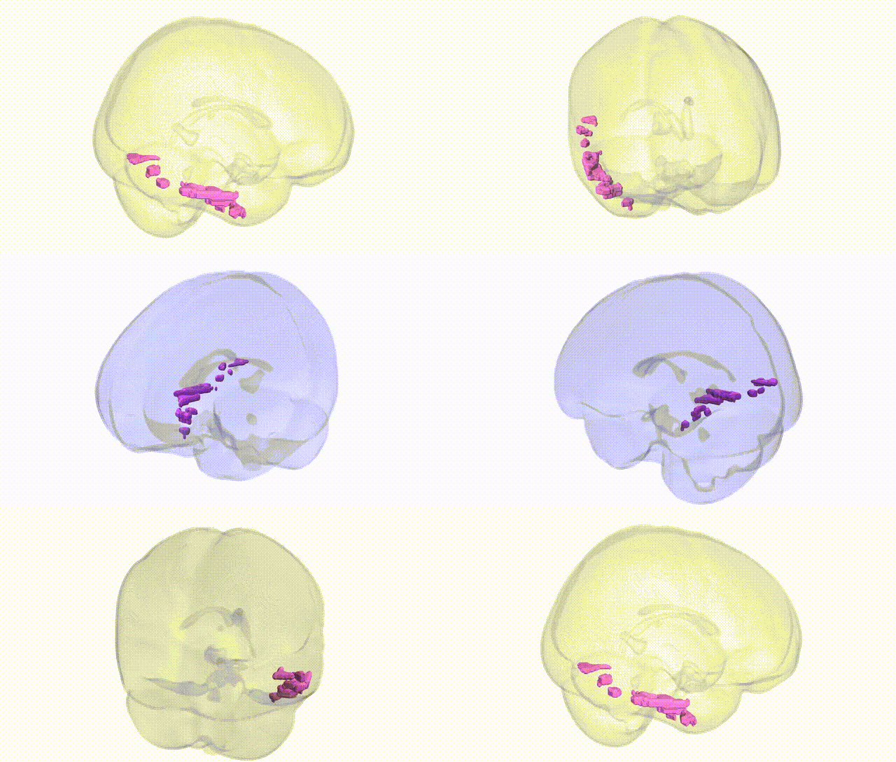
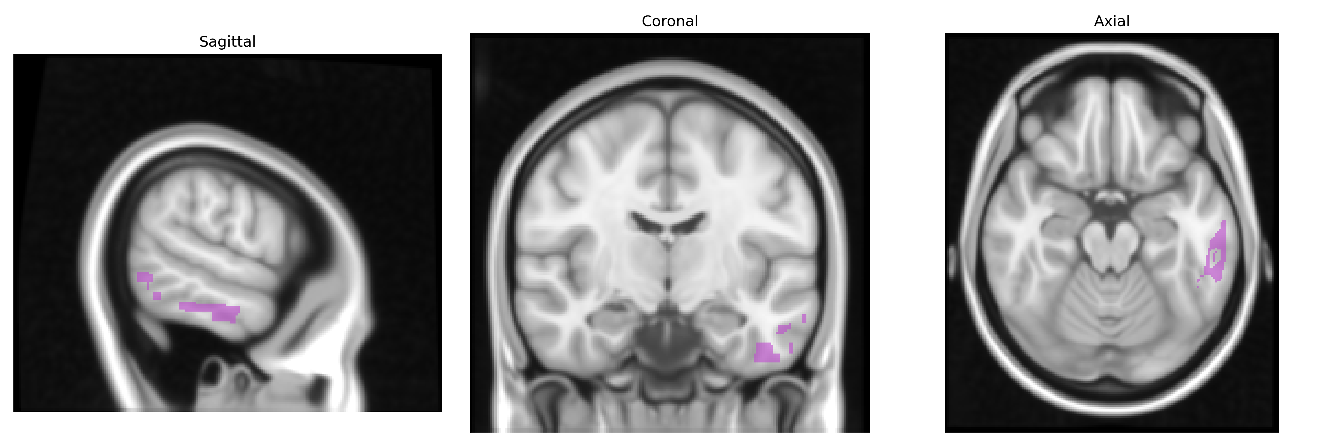
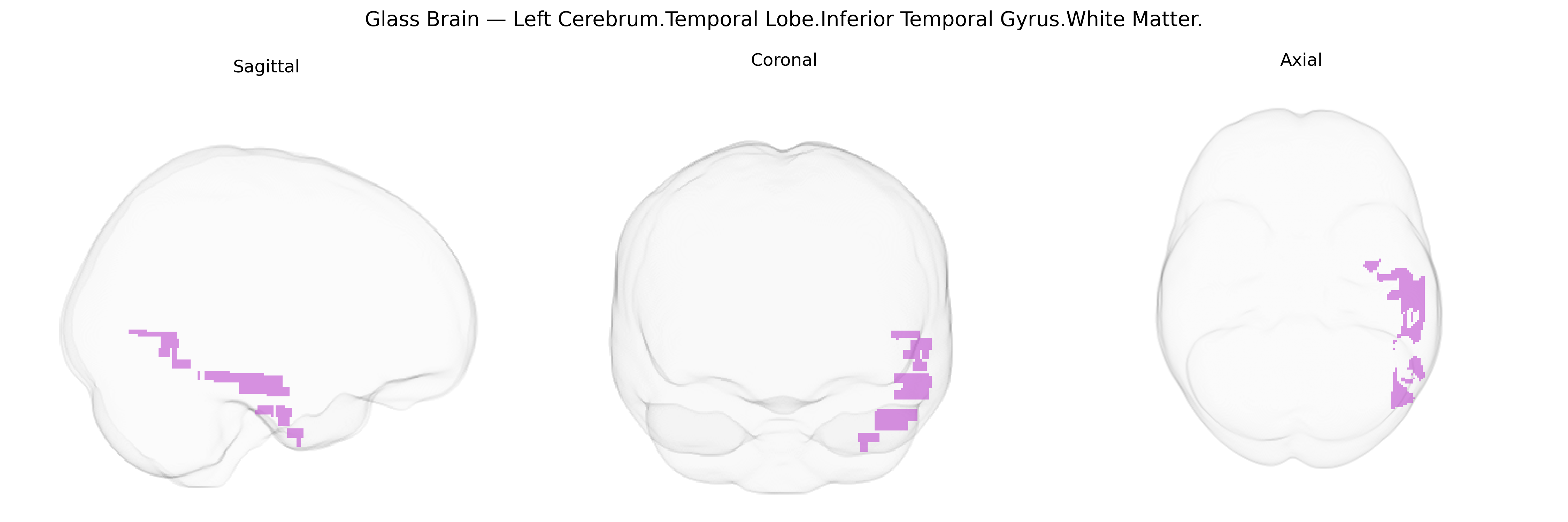
The bilateral Left Cerebrum.Temporal Lobe.Inferior Temporal Gyrus.White Matter corresponds to the subcortical fiber systems underlying the inferior temporal gyrus in the left temporal lobe, including components of long-range association pathways such as portions of the inferior longitudinal fasciculus and other ventral stream tracts that connect occipital visual areas with anterior temporal and frontal regions. Functionally, this white matter territory supports the transmission of visual object and form information crucial for high-level visual recognition, semantic processing, and aspects of language and memory that rely on ventral temporal networks. Lesions in this region can disrupt visual object recognition and contribute to neurocognitive syndromes such as visual agnosia or impairments in semantic memory. There is no direct link for the inferior temporal gyrus white matter; a related cortical structure is the Inferior temporal gyrus.
The bilateral Left Cerebrum.Temporal Lobe.Inferior Temporal Gyrus.White Matter, as defined in the Talairach 1 mm atlas, lies along major ventral visual and associative pathways (including portions of the inferior longitudinal fasciculus and adjacent tracts), and genetic associations involving this region derive largely from imaging–genetics and GWAS of white-matter microstructure, cortical connectivity, and neurodevelopmental or neurodegenerative disorders rather than region-specific candidate-gene studies. Large-scale diffusion MRI GWAS (for example, UK Biobank–based studies of fractional anisotropy and mean diffusivity in temporal association fibers) have identified polygenic influences enriched for genes regulating axon guidance, myelination, and synaptic organization (such as variants near NTRK2, CNTN4, NRXN1, and multiple oligodendrocyte-related loci), implicating this white matter in general neurodevelopmental wiring and visual–semantic networks. Indirectly, genetic risk for autism spectrum disorder, schizophrenia, and major depressive disorder—captured by polygenic risk scores—has been associated with altered integrity or connectivity in inferior temporal and adjacent ventral-stream white matter, while Alzheimer’s disease and frontotemporal dementia risk loci (including APOE and MAPT haplotypes) have been linked to degeneration or reduced structural connectivity in temporal lobe association pathways that traverse or border this region. GWAS of face recognition, object recognition, and reading ability/rapid automatized naming, which functionally recruit inferior temporal cortex, also show polygenic influences overlapping neurodevelopmental and synaptic genes, though specific single variants uniquely targeting this precise Talairach white-matter label have not been robustly isolated, and current evidence supports a highly polygenic, distributed genetic architecture rather than discrete region-specific genetic effects.
Overview generated by GPT-4o (2026).
Region ID: 14
Hemisphere: bilateral
Atlas: Talairach labels 1mm
Full Quality Version: Download MP4
Full Quality Version: Download MP4
Wali Sidiqyar*, Gaurav Rudravaram*, Elyssa M. McMaster, Trent M. Schwartz, Adam M. Saunders, Kurt G. Schilling, Bennett A. Landman "Introducing SPINS: A Shared Public Visualization Library of Neuroanatomical Structures." Medical Imaging with Deep Learning- short paper
This resource is licensed under CC0 1.0 Universal (Public Domain).