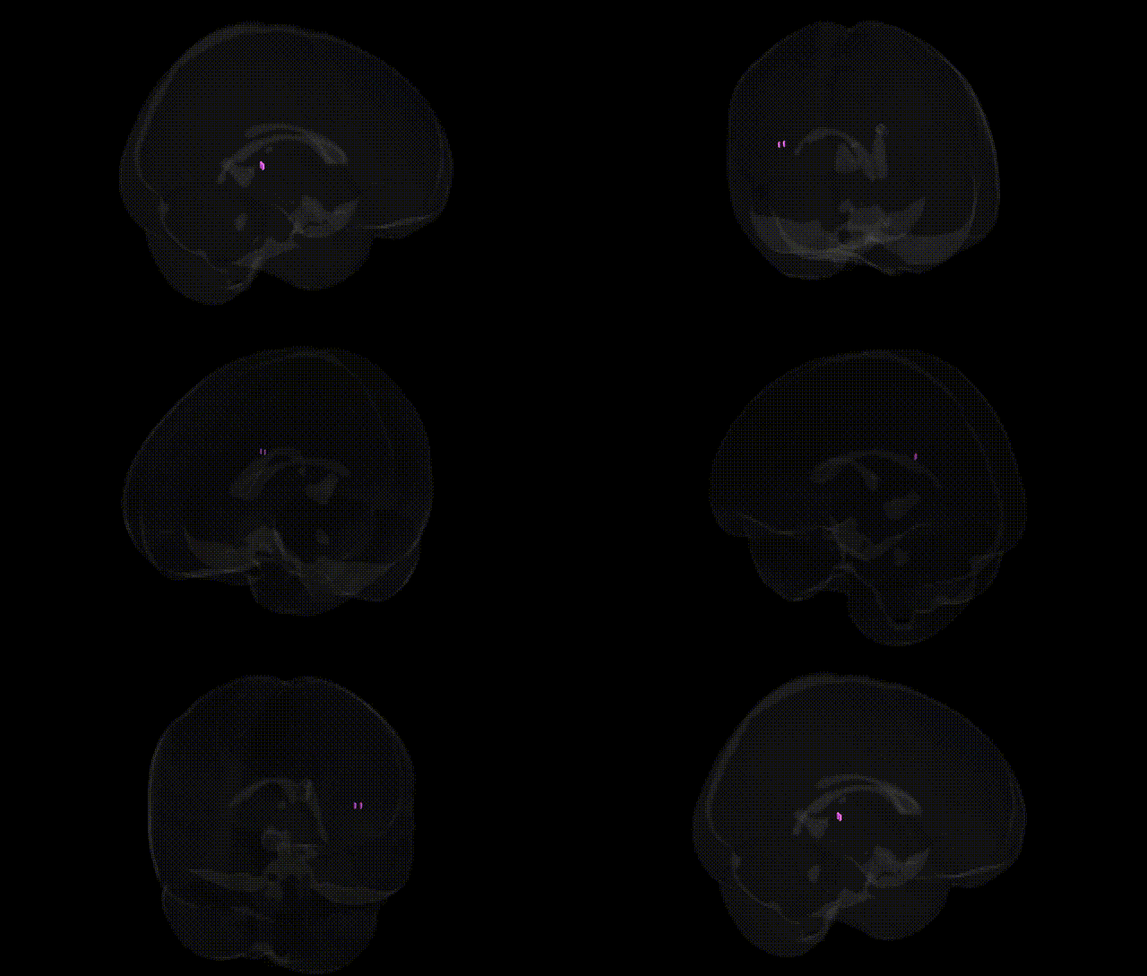
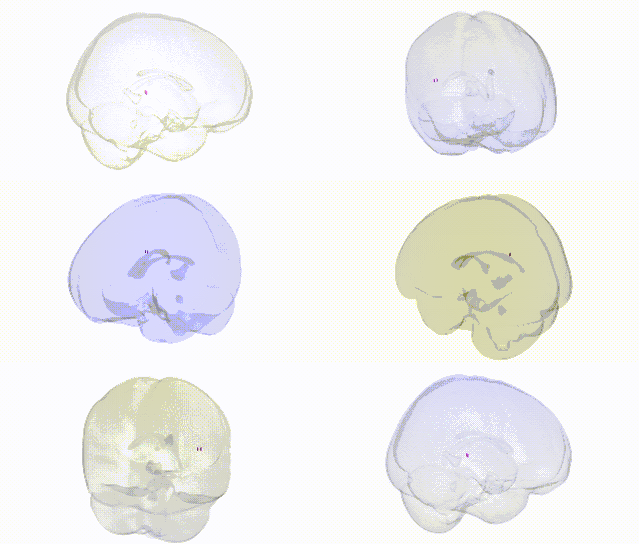
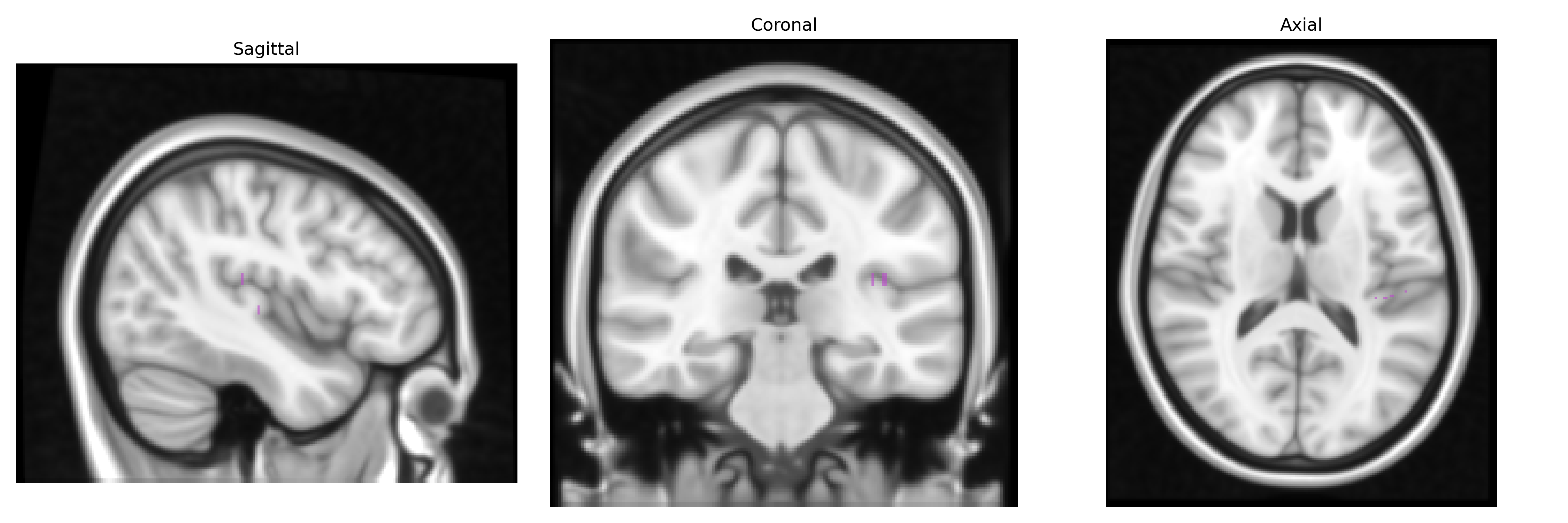
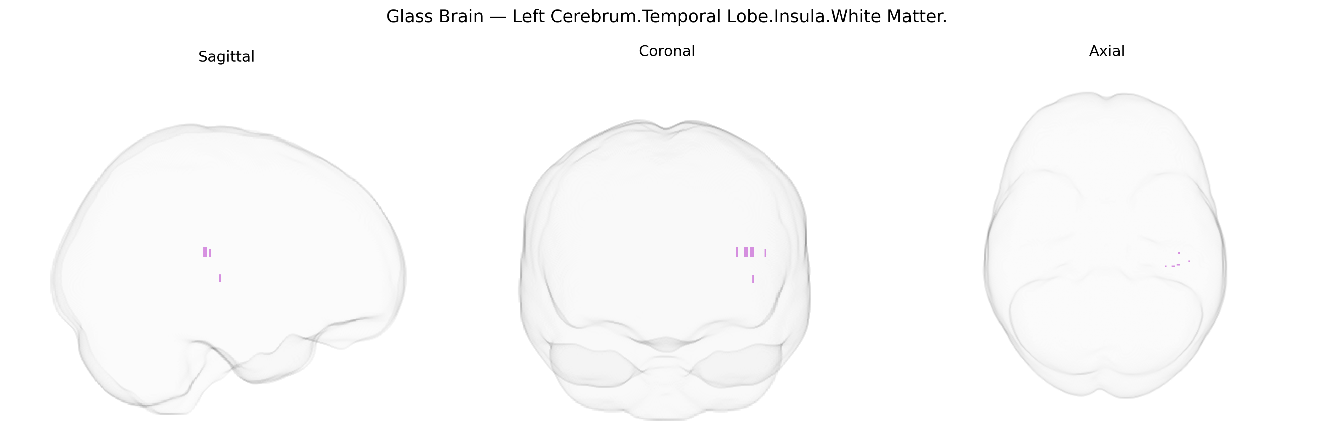
The bilateral Left Cerebrum.Temporal Lobe.Insula.White Matter corresponds to subcortical fiber pathways coursing through the white matter of the insular region within the temporal lobe of the left cerebral hemisphere, as defined by the Talairach 1 mm atlas. This area contains association, commissural, and projection fibers that interconnect insular cortex with temporal, frontal, and parietal regions, contributing to networks involved in multimodal sensory integration, visceral-autonomic processing, emotional salience, and aspects of language and interoception. Although the white matter itself lacks a dedicated article, it lies deep to and supports the function of the overlying insular cortex. There is no direct link for this exact white matter label; a closely related structure is the Insular cortex.
The bilateral left temporal insular white matter lies within frontotemporal–limbic networks whose microstructure and volume show modest but replicable SNP-based heritability in large imaging-genetics cohorts (e.g., ENIGMA, UK Biobank), with common variation in genes involved in axonal guidance, myelination, and synaptic signaling (such as NTRK1/2, CNTN4/6, LINGO1, and various oligodendrocyte-related loci) contributing to inter-individual differences in white matter integrity. GWAS of diffusion tensor imaging measures have identified associations between insula-adjacent temporal white matter tracts and polygenic risk scores for major depressive disorder, schizophrenia, bipolar disorder, and autism spectrum disorder, consistent with the insula’s role in salience, interoception, and affective regulation. Reduced fractional anisotropy and altered connectivity in this region are genetically correlated with higher liability to psychosis and mood disorders, and several studies report overlap between insular white matter–associated loci and those implicated in neurodevelopmental traits, cognitive performance, pain sensitivity, and substance use, although no single variant shows a uniquely specific, high-effect association with this precise Talairach-defined subregion.
Overview generated by GPT-4o (2026).
Region ID: 635
Hemisphere: bilateral
Atlas: Talairach labels 1mm
Full Quality Version: Download MP4
Full Quality Version: Download MP4
Wali Sidiqyar*, Gaurav Rudravaram*, Elyssa M. McMaster, Trent M. Schwartz, Adam M. Saunders, Kurt G. Schilling, Bennett A. Landman "Introducing SPINS: A Shared Public Visualization Library of Neuroanatomical Structures." Medical Imaging with Deep Learning- short paper
This resource is licensed under CC0 1.0 Universal (Public Domain).