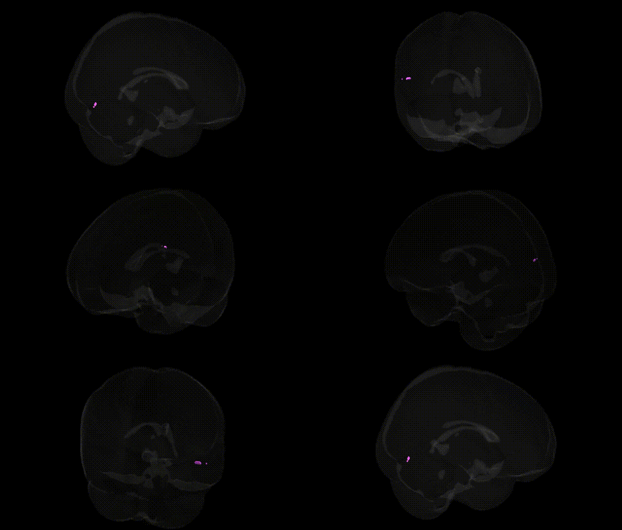
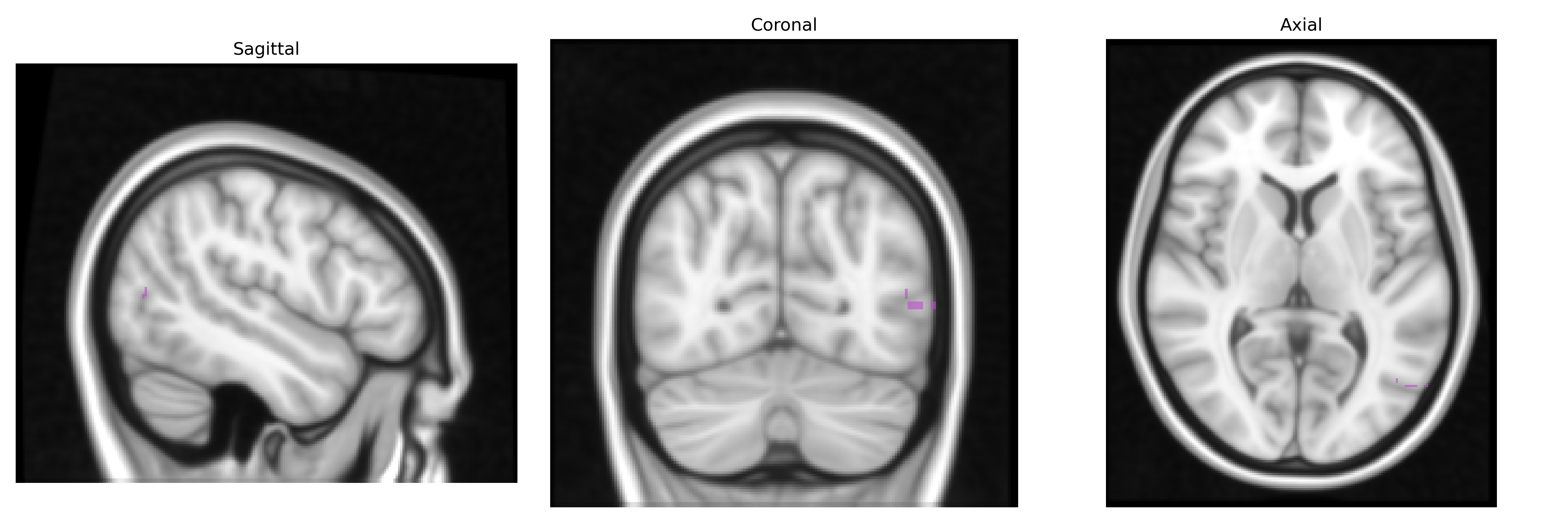
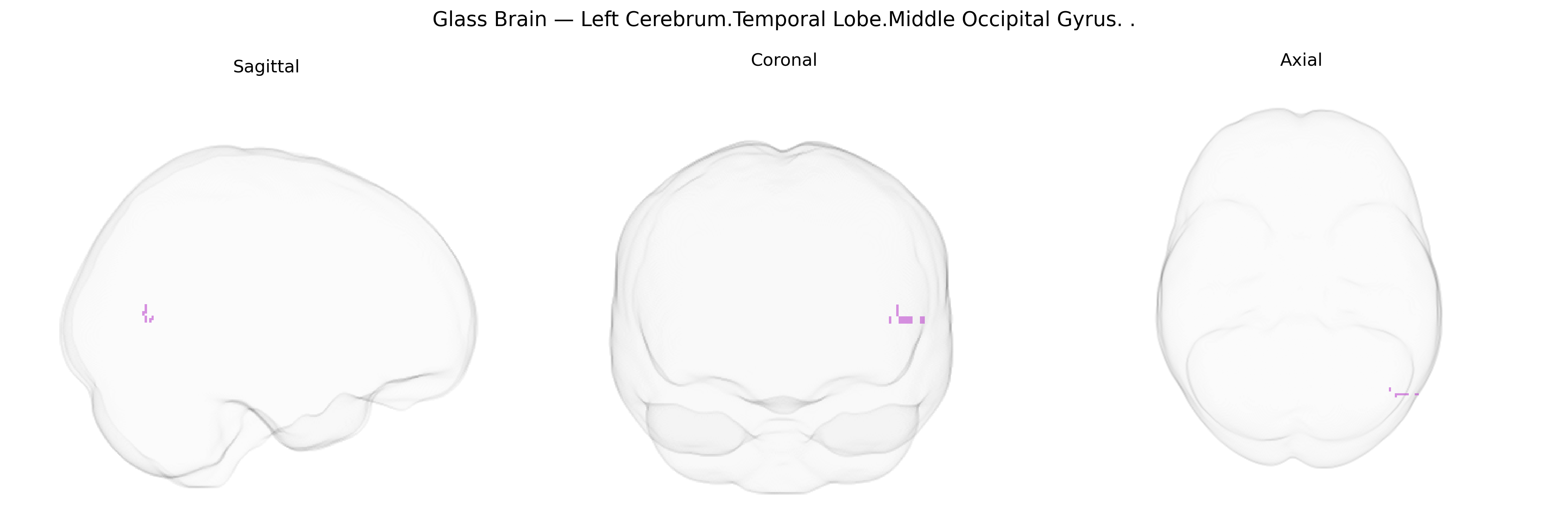
The bilateral Left Cerebrum.Temporal Lobe.Middle Occipital Gyrus in the Talairach 1 mm Atlas refers to voxels in the left cerebral hemisphere that are labeled at the intersection of the temporal lobe and the middle portion of the occipital gyrus, a region generally corresponding to parts of the lateral occipital cortex involved in mid‑ to high‑level visual processing. Functionally, this area participates in processing visual form, motion, and object features, and often overlaps with regions engaged in visual association, such as components of the ventral and dorsal visual streams. In many neuroimaging studies, this label captures portions of cortex bordering or overlapping with areas like V3/V4 and lateral occipital complex, implicated in shape, object, and scene perception as well as integration of visual information for recognition and spatial analysis. There is no direct Wikipedia article for this exact Talairach label; a related structure is the Lateral occipital cortex.
The Middle Occipital Gyrus (MOG) of the temporal-occipital region, as localized in the Talairach 1 mm atlas, has been implicated in multiple genetic and GWAS-based associations through its role in visual processing, higher-order perception, and large-scale functional networks rather than via region-specific genes. Common variants in synaptic and neurodevelopmental genes such as BDNF, NRG1, DISC1, and CACNA1C, repeatedly linked to schizophrenia, bipolar disorder, and major depressive disorder, have been associated with altered occipital and temporo-occipital cortical thickness or volume in imaging-genetics studies, including bilateral MOG involvement in case–control and endophenotype analyses. GWAS of cortical thickness and surface area (e.g., ENIGMA and UK Biobank–based consortia) have identified loci in genes related to neuronal migration, axon guidance, and myelination (such as HRK, MIR124-1, WNT pathway genes, and multiple intergenic regulatory regions) that influence morphology of occipital association cortex, with signals overlapping the middle occipital and lateral temporal areas. Polygenic risk scores for schizophrenia, autism spectrum disorder, and ADHD show associations with altered activation or structure in the MOG during tasks involving visual attention, motion perception, and social cognition, reflecting a convergence between genetic risk for neurodevelopmental disorders and functional alterations in this region. Moreover, GWAS of visual acuity, refractive error (myopia), and visual processing speed have reported variants near genes involved in phototransduction, retinal development, and cortical visual pathways that correlate with functional and structural measures in occipital cortex, including the MOG. Migraine and visual aura GWAS, implicating genes in glutamatergic signaling and vascular regulation (such as TRPM8, LRP1, and others), also point to altered occipital excitability and perfusion patterns that encompass the middle occipital gyrus. Overall, existing evidence suggests that genetic effects on this bilateral temporal–occipital region are largely mediated by polygenic influences on cortical development, synaptic plasticity, and neurovascular function, rather than any single gene uniquely linked to the Middle Occipital Gyrus.
Overview generated by GPT-4o (2026).
Region ID: 625
Hemisphere: bilateral
Atlas: Talairach labels 1mm
Full Quality Version: Download MP4
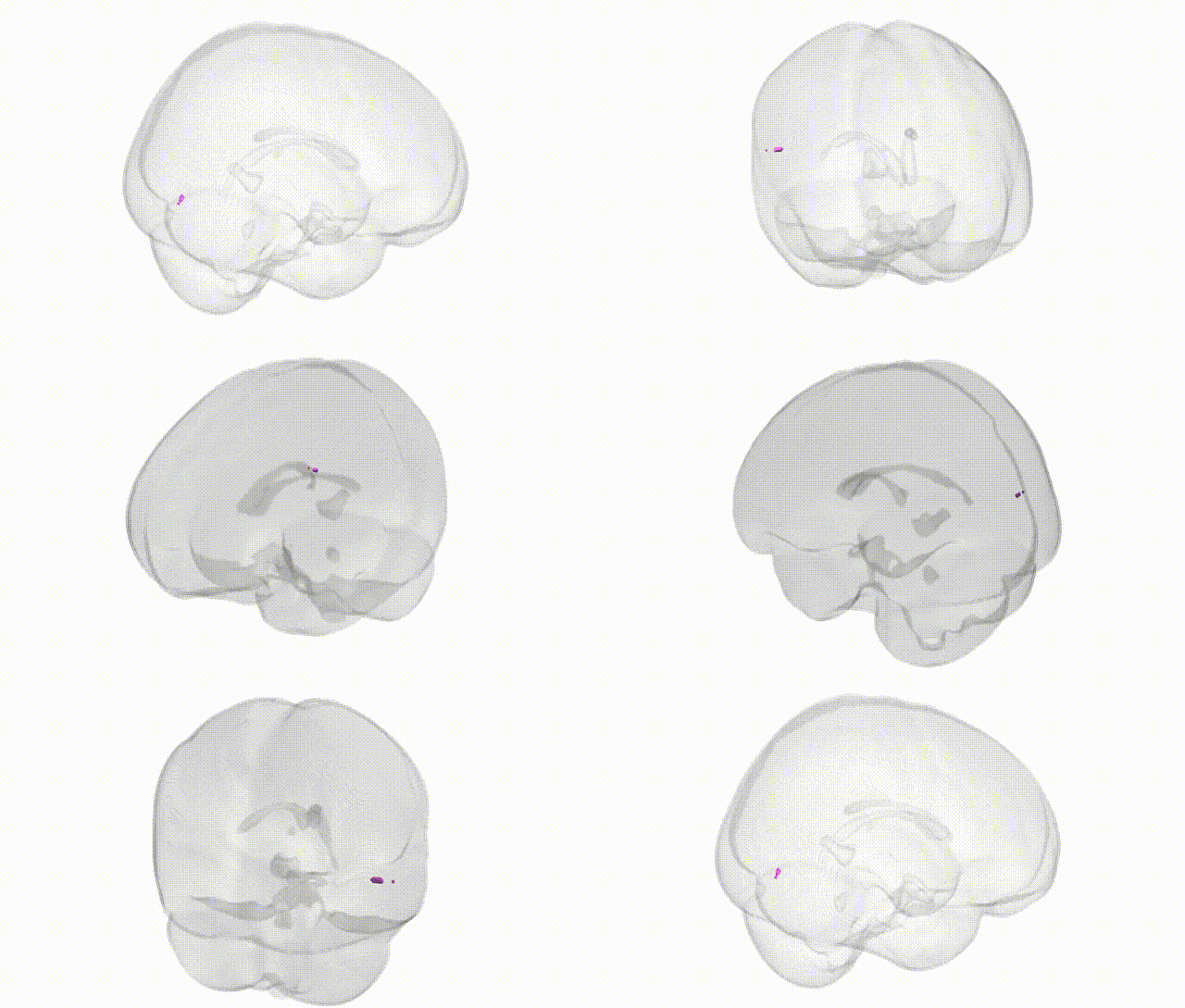
Full Quality Version: Download MP4
Wali Sidiqyar*, Gaurav Rudravaram*, Elyssa M. McMaster, Trent M. Schwartz, Adam M. Saunders, Kurt G. Schilling, Bennett A. Landman "Introducing SPINS: A Shared Public Visualization Library of Neuroanatomical Structures." Medical Imaging with Deep Learning- short paper
This resource is licensed under CC0 1.0 Universal (Public Domain).