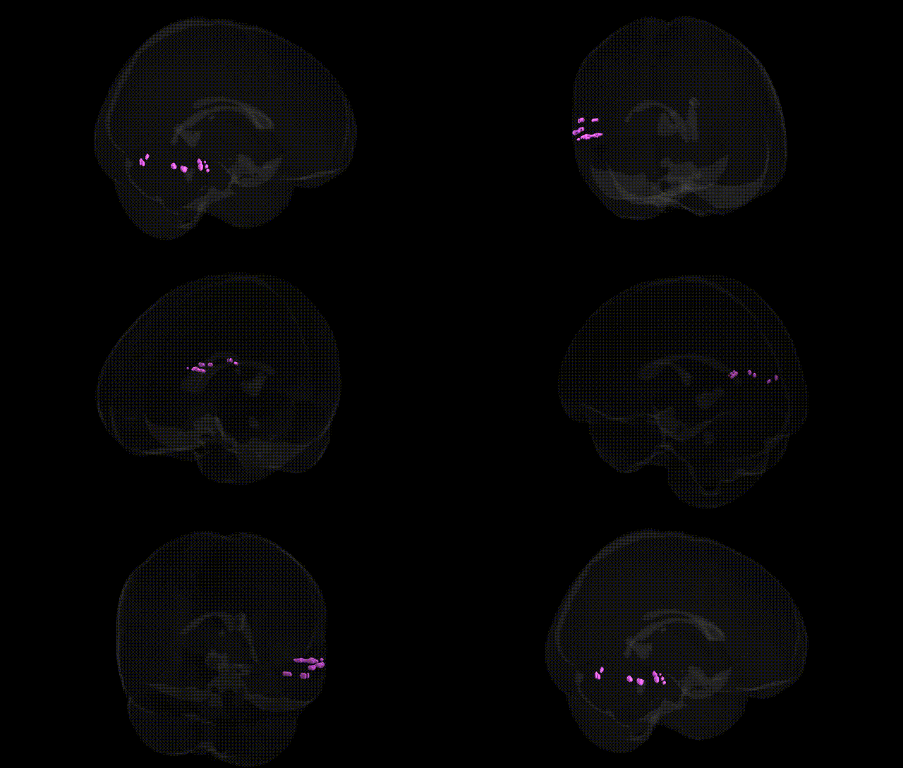
The bilateral left cerebrum temporal lobe middle temporal gyrus gray matter corresponds to cortical tissue located on the lateral aspect of the temporal lobe, between the superior and inferior temporal gyri, and extending along the anteroposterior axis from the temporal pole toward the temporo-occipital junction. This region is implicated in higher-order auditory and language processing (particularly lexical-semantic retrieval), visual motion and object recognition, and aspects of social cognition such as theory of mind and processing of biological motion. Cytoarchitectonically, it encompasses portions of Brodmann areas (e.g., BA21 and adjacent fields), with extensive connectivity to the superior temporal sulcus, inferior parietal lobule, prefrontal cortex, and limbic structures, thereby participating in distributed networks supporting multimodal integration and semantic memory. No direct link exists for “middle temporal gyrus gray matter” as a standalone entry; a related structure is the Middle temporal gyrus.
The bilateral middle temporal gyrus gray matter in the left cerebrum has been implicated in multiple genetic and GWAS-based associations, particularly involving language, semantic processing, social cognition, and psychiatric vulnerability. Large-scale imaging–genetics consortia such as ENIGMA and UK Biobank have identified common variants near genes involved in neurodevelopment and synaptic function (e.g., MAPT, BDNF, GRIN2B, DLX and FOXP family genes) that influence cortical thickness and surface area in temporal lobe regions encompassing the middle temporal gyrus, with several loci reaching genome-wide significance for temporal lobe volume and cortical metrics. GWAS of schizophrenia, bipolar disorder, and major depressive disorder have reported risk alleles that correlate with structural and functional alterations in the middle temporal gyrus, while autism spectrum disorder and language-related traits (including reading ability and developmental language disorder) show associations between variants in language and axon-guidance genes (such as CNTNAP2 and ROBO/SLIT pathways) and atypical middle temporal gyrus morphology or connectivity. Alzheimer’s disease and other dementias, particularly via APOE ε4 and loci involved in amyloid and tau pathways, have been linked to early atrophy and hypometabolism in the middle temporal gyrus, highlighting a genetic contribution to temporal lobe degeneration. Together, these findings indicate that common and rare variants affecting synaptic plasticity, neurodevelopment, and neurodegeneration contribute to individual differences and disease-related changes in the left middle temporal gyrus as defined in Talairach 1 mm atlas parcellations.
Overview generated by GPT-4o (2026).
Region ID: 528
Hemisphere: bilateral
Atlas: Talairach labels 1mm
Full Quality Version: Download MP4
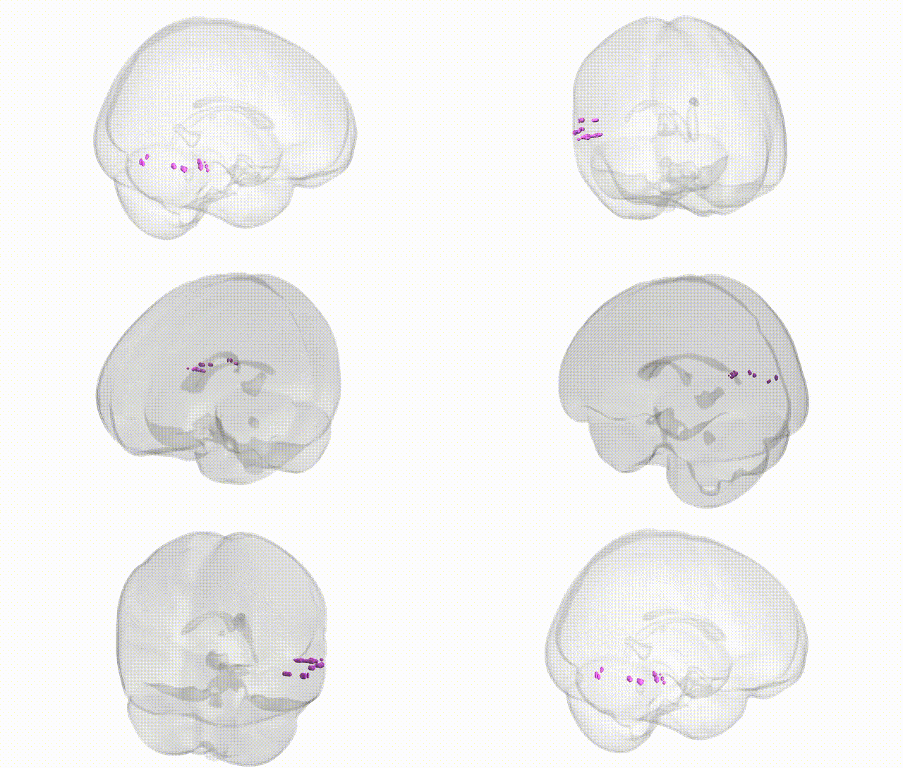
Full Quality Version: Download MP4
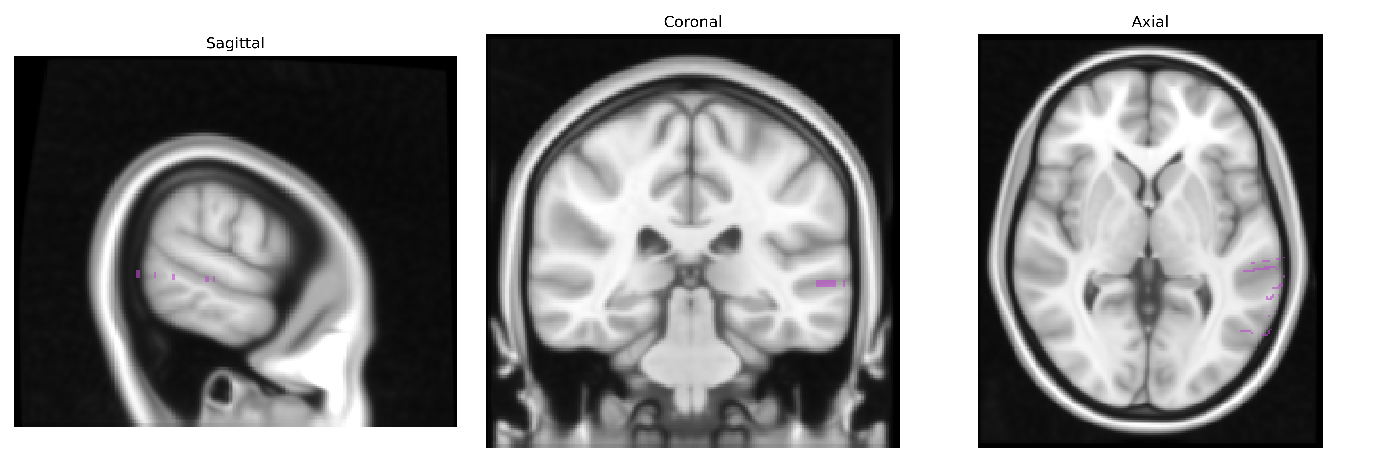
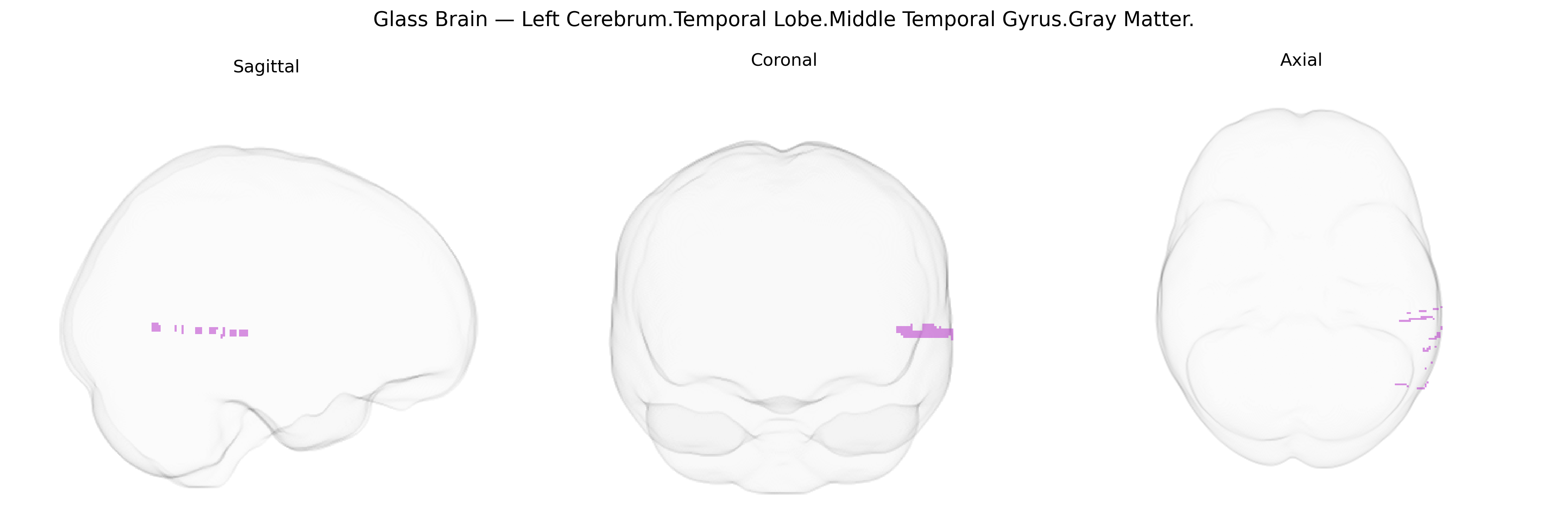
Wali Sidiqyar*, Gaurav Rudravaram*, Elyssa M. McMaster, Trent M. Schwartz, Adam M. Saunders, Kurt G. Schilling, Bennett A. Landman "Introducing SPINS: A Shared Public Visualization Library of Neuroanatomical Structures." Medical Imaging with Deep Learning- short paper
This resource is licensed under CC0 1.0 Universal (Public Domain).