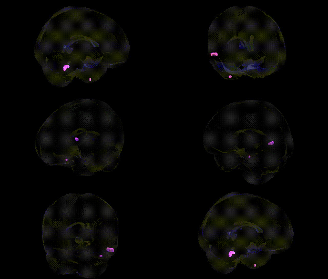
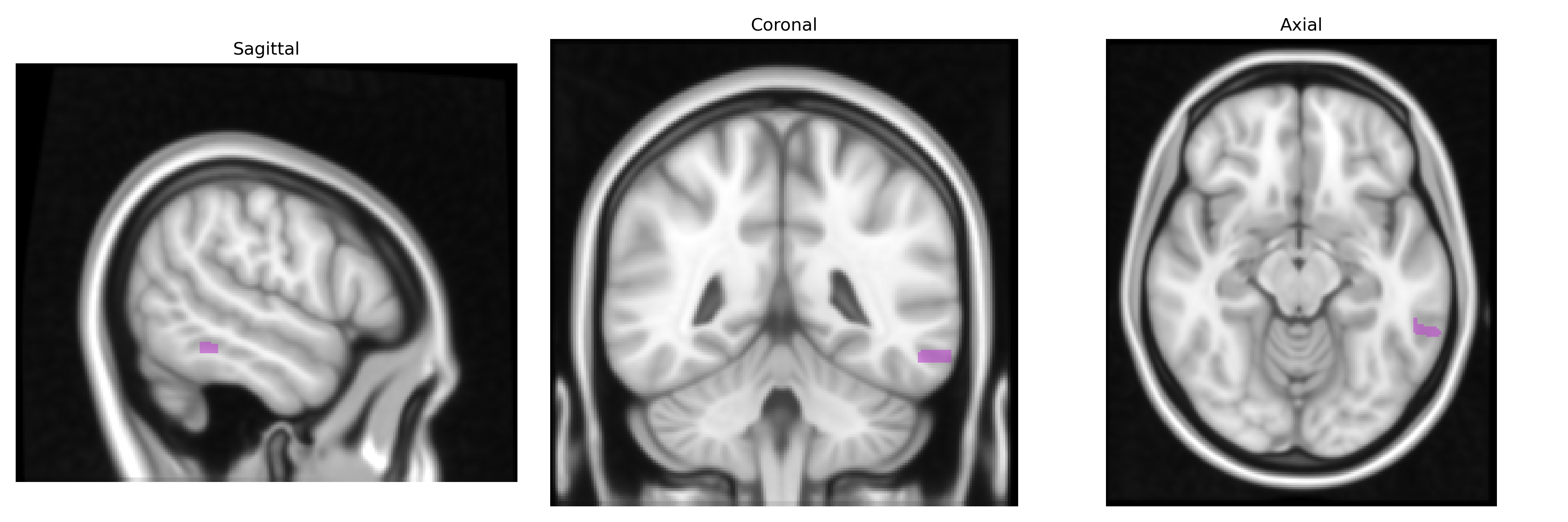
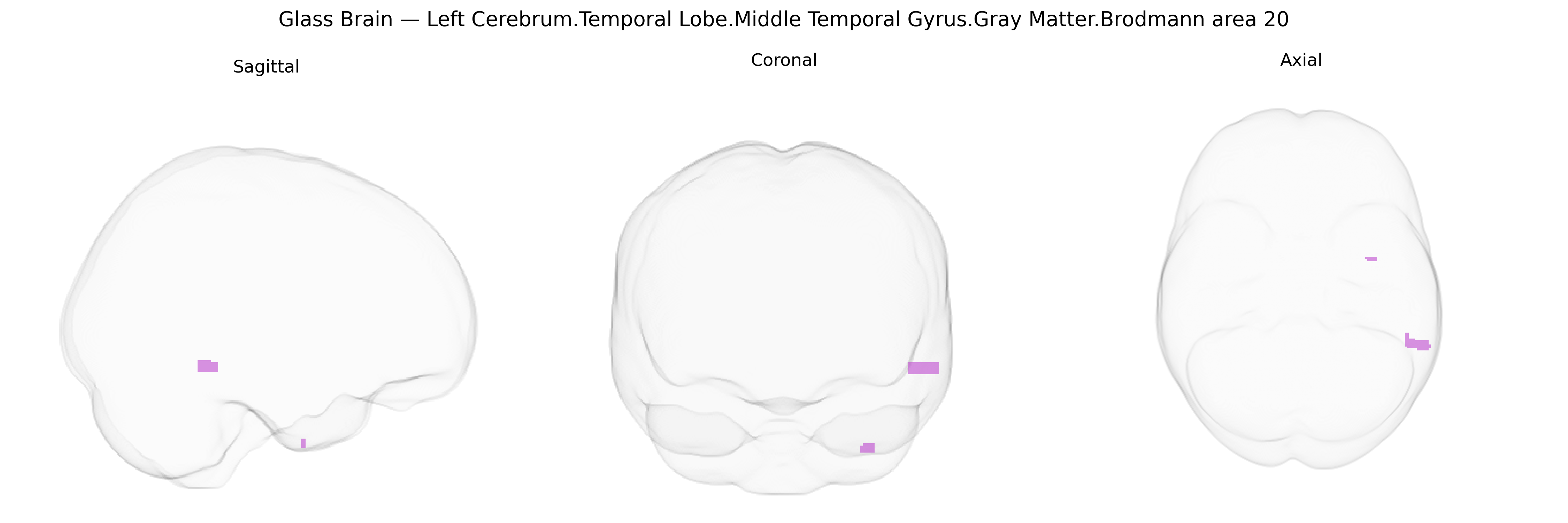
The bilateral Left Cerebrum.Temporal Lobe.Middle Temporal Gyrus.Gray Matter.Brodmann area 20 corresponds to a ventral portion of the temporal neocortex located on the inferolateral aspect of the temporal lobe, anterior to the occipitotemporal regions and inferior to the superior temporal sulcus. Histologically defined by its granular cortex and association-type lamination, Brodmann area 20 participates in high-level visual object processing, including face and complex shape recognition, and contributes to semantic memory, lexical access, and integration of multimodal sensory information. Functional connectivity links this region with fusiform, inferior temporal, and prefrontal association areas, supporting roles in conceptual knowledge, language-related processes, and context-dependent perception. Lesions in this area can result in deficits such as visual agnosia or impairments in semantic processing, reflecting its importance as a convergence zone in the ventral visual and temporal association networks. No direct article for Brodmann area 20 in the middle temporal gyrus exists; a related entry is Middle temporal gyrus.
The bilateral middle temporal gyrus (MTG) in Brodmann area 20, as defined in the Talairach 1 mm atlas, has been implicated in multiple genetic and genome‑wide association studies that link its gray matter structure and function to diverse neuropsychiatric and cognitive phenotypes. GWAS and imaging‑genetics consortia (e.g., ENIGMA) have reported associations between MTG cortical thickness or surface area and common variants in genes involved in neurodevelopment, synaptic function, and axonal guidance (such as BDNF, NRG1, DISC1, CNTNAP2, and genes regulating glutamatergic and GABAergic signaling), although effects are typically small and distributed across the cortex. Structural and functional alterations in BA20/MTG show heritability and have been associated with polygenic risk scores for schizophrenia, major depression, autism spectrum disorder, and Alzheimer’s disease, with some studies linking APOE, CLU, and other AD‑risk loci to temporal lobe atrophy patterns that include the MTG. Genetic variants influencing language and semantic processing (e.g., FOXP2‑related pathways and CNTNAP2) and reading or dyslexia risk loci have also been connected to activation and morphology in temporal association cortices encompassing BA20. Additionally, common variants associated with general cognitive ability, educational attainment, and social cognition have been correlated with MTG volume or connectivity in large‑scale imaging‑GWAS, suggesting that BA20 serves as a convergent site where polygenic influences on higher‑order visual, semantic, and socio‑emotional processing manifest at the level of gray matter architecture and function, even though no single gene shows a specific, exclusive association with this region.
Overview generated by GPT-4o (2026).
Region ID: 23
Hemisphere: bilateral
Atlas: Talairach labels 1mm
Full Quality Version: Download MP4
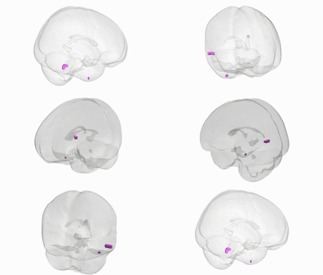
Full Quality Version: Download MP4
Wali Sidiqyar*, Gaurav Rudravaram*, Elyssa M. McMaster, Trent M. Schwartz, Adam M. Saunders, Kurt G. Schilling, Bennett A. Landman "Introducing SPINS: A Shared Public Visualization Library of Neuroanatomical Structures." Medical Imaging with Deep Learning- short paper
This resource is licensed under CC0 1.0 Universal (Public Domain).