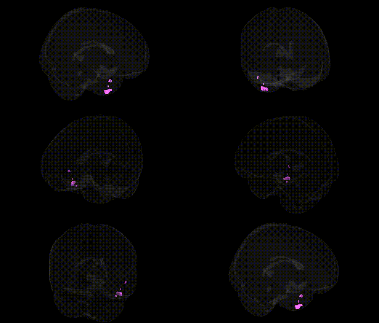
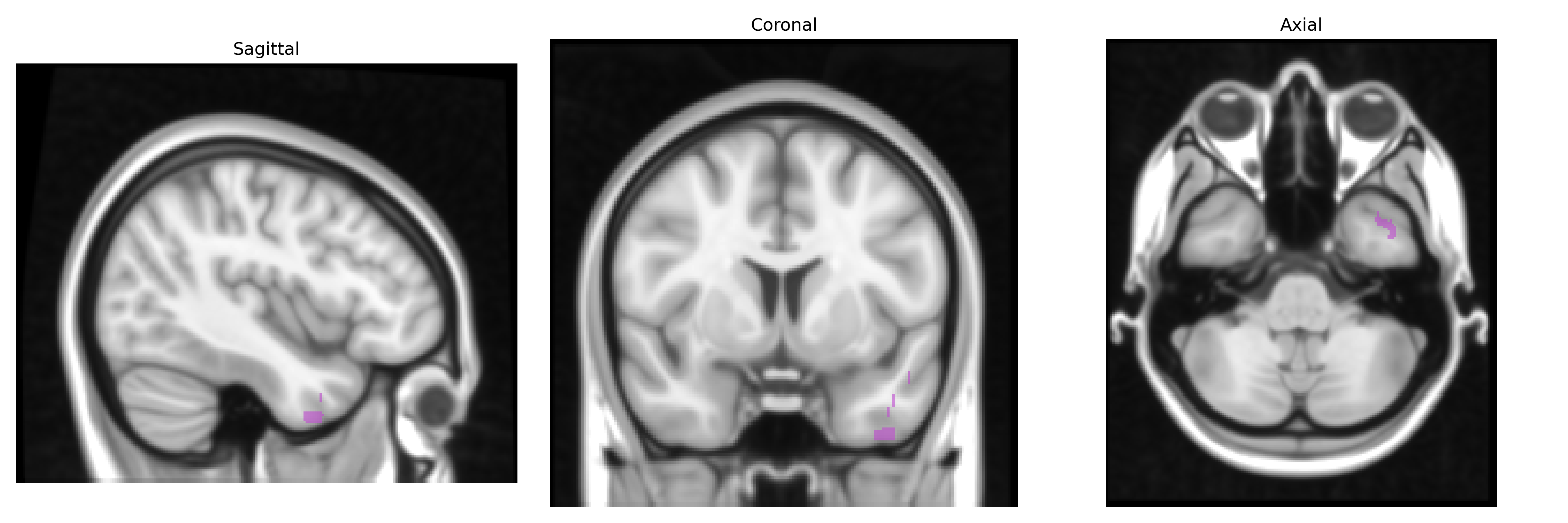
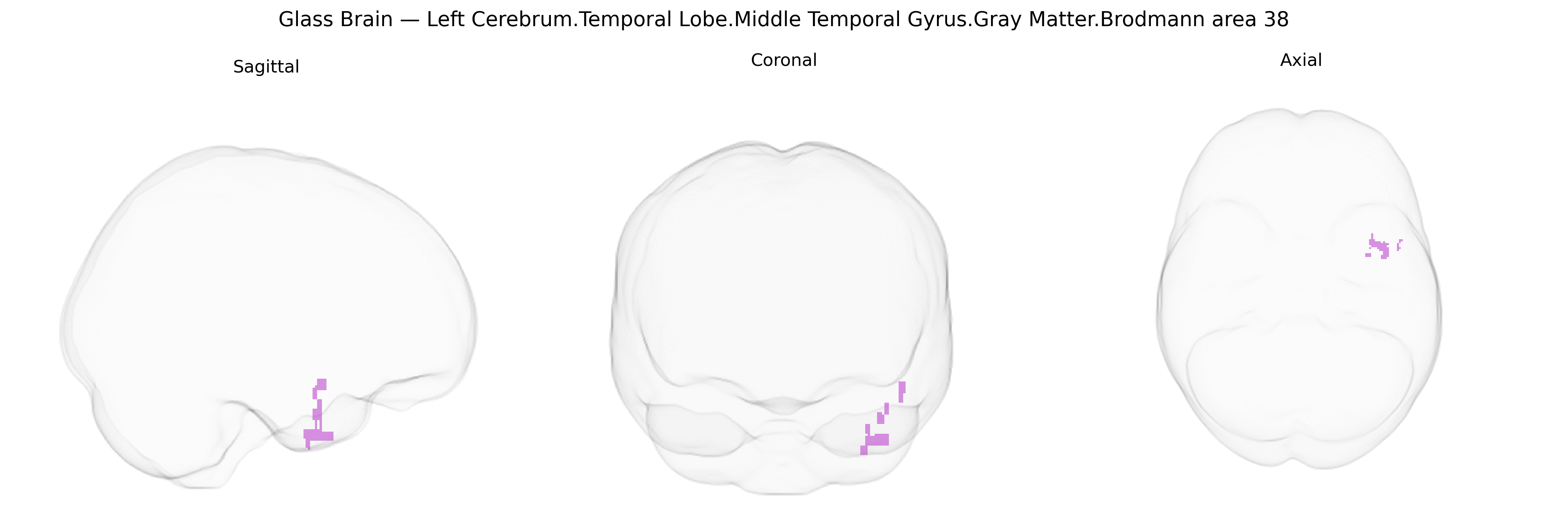
The bilateral Left Cerebrum.Temporal Lobe.Middle Temporal Gyrus.Gray Matter.Brodmann area 38 corresponds to the most anterior portion of the temporal lobe, commonly referred to as the temporal pole, and is composed of cortical gray matter within Brodmann area 38. This region participates in higher-order multimodal association processes, including semantic memory, social and emotional cognition, and integration of complex auditory and visual information with affective and contextual cues. It maintains extensive reciprocal connections with limbic and paralimbic structures (such as the amygdala, orbitofrontal cortex, and hippocampal formation) and with other temporal and frontal association areas, supporting roles in language-related semantic processing, person and face recognition, and emotional valence assignment. Dysfunction or structural pathology in this region, particularly in the left hemisphere, has been associated with semantic variant primary progressive aphasia and other temporal lobe neurodegenerative and epileptogenic conditions. Brodmann area 38
The bilateral left temporal pole (middle temporal gyrus, Brodmann area 38) shows convergent genetic associations from imaging-genetics and GWAS studies implicating neurodevelopmental, synaptic, and psychiatric risk variants: ENIGMA and other large consortia have reported heritable variation in temporal pole cortical thickness and surface area linked to common variants in genes such as HMGA2, IGF1, and other growth‐ and neurodevelopment-related loci, while polygenic scores for schizophrenia, bipolar disorder, major depression, and autism spectrum disorder associate with structural and functional alterations in this region. Temporal pole volume and connectivity have been implicated in GWAS of temporal lobe epilepsy—where genes like SCN1A, GABRA2, and other ion channel and synaptic genes contribute to seizure susceptibility—and in frontotemporal dementia and semantic variant primary progressive aphasia, in which mutations in MAPT, GRN, C9orf72, and related genes produce early and selective atrophy of anterior temporal cortices including BA38. Additional GWAS of social cognition, face processing, language abilities, and personality traits (e.g., extraversion, neuroticism) identify polygenic influences that overlap with networks centered on or strongly involving the temporal pole, supporting a role for genetically mediated variation in this region in semantic memory, socioemotional processing, and vulnerability to neuropsychiatric and neurodegenerative disorders.
Overview generated by GPT-4o (2026).
Region ID: 33
Hemisphere: bilateral
Atlas: Talairach labels 1mm
Full Quality Version: Download MP4
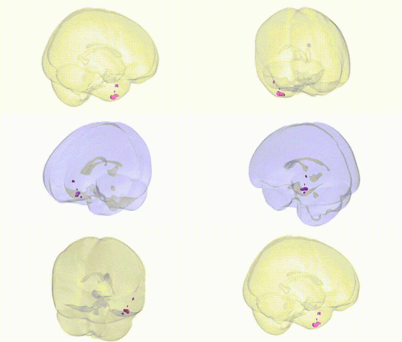
Full Quality Version: Download MP4
Wali Sidiqyar*, Gaurav Rudravaram*, Elyssa M. McMaster, Trent M. Schwartz, Adam M. Saunders, Kurt G. Schilling, Bennett A. Landman "Introducing SPINS: A Shared Public Visualization Library of Neuroanatomical Structures." Medical Imaging with Deep Learning- short paper
This resource is licensed under CC0 1.0 Universal (Public Domain).