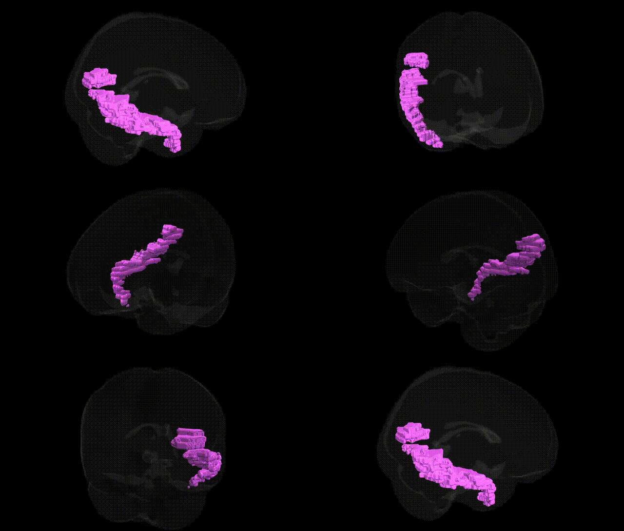
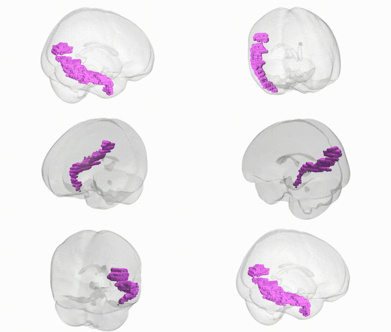
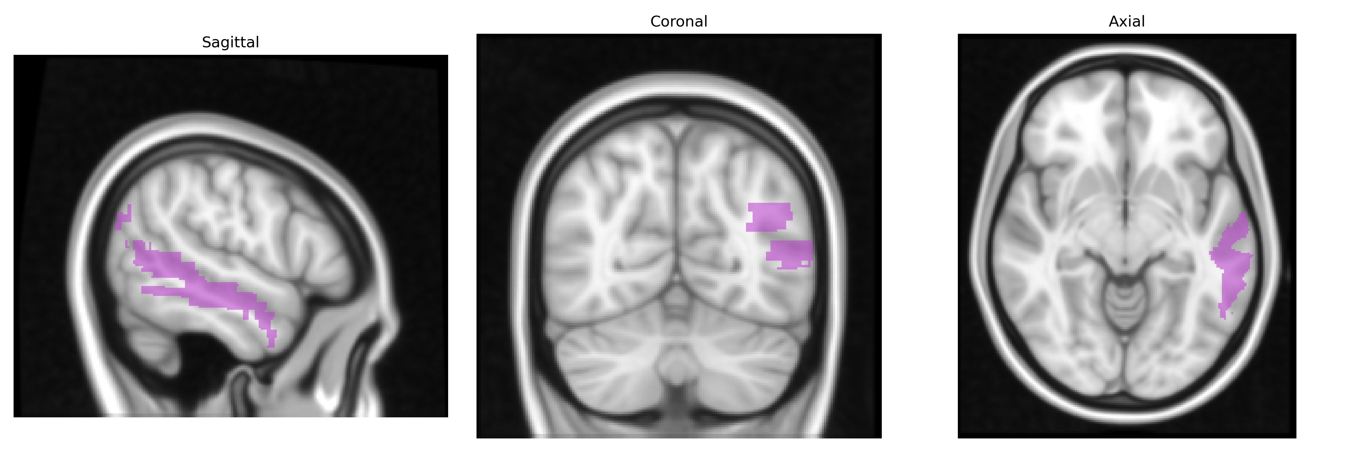
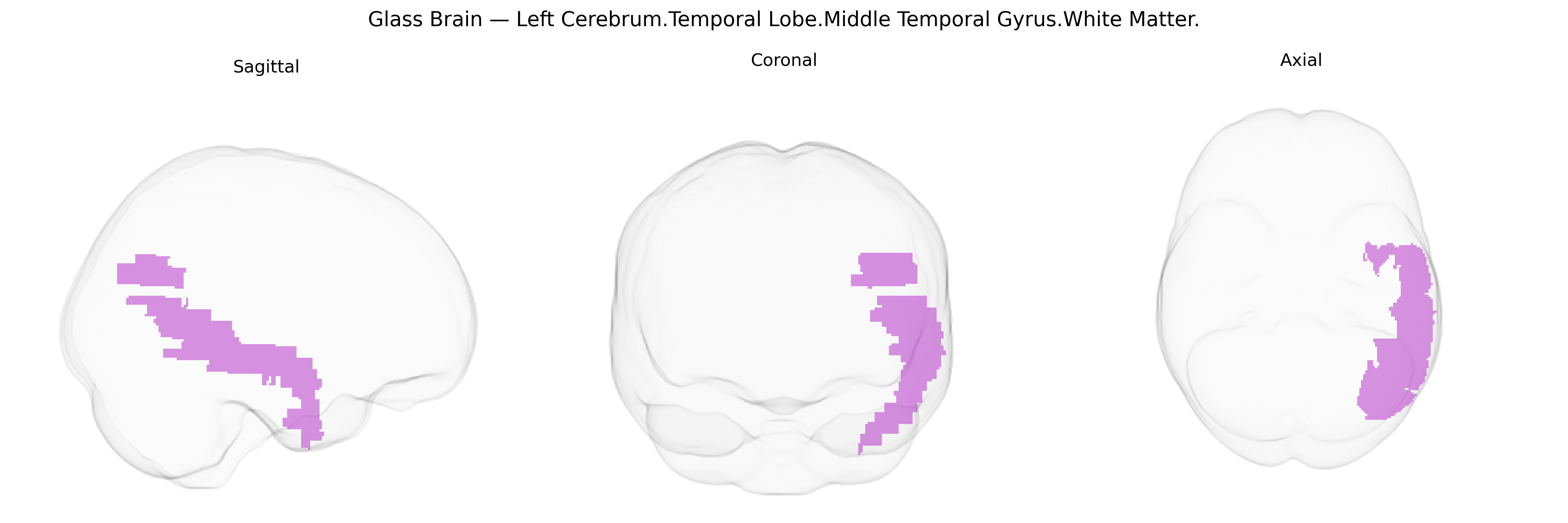
The bilateral Left Cerebrum.Temporal Lobe.Middle Temporal Gyrus.White Matter corresponds to the subcortical myelinated fiber pathways coursing beneath the middle temporal gyrus in the temporal lobe of the left cerebral hemisphere, as defined in the Talairach 1 mm atlas. This white matter region contains association fibers interconnecting temporal neocortical areas (including middle and inferior temporal gyri), as well as pathways linking temporal regions with occipital, parietal, and frontal cortices, thereby contributing to multimodal sensory integration, language processing, semantic memory, and higher-order visual recognition. It also carries portions of major fasciculi such as the inferior longitudinal fasciculus and arcuate/longitudinal association pathways that support communication between temporal lobe structures and distributed cortical networks. There is no direct link for this specific white matter label; a related cortical structure is the Middle temporal gyrus.
The bilateral white matter of the left middle temporal gyrus (MTG) has been implicated in several genetic and genome-wide association studies via imaging-genetics and voxel-based morphometry, although few findings are region-exclusive. Variants in genes affecting myelination and white matter integrity, such as those in the CNTNAP2, NRG1, and ERBB4 pathways, have been associated with altered temporal lobe white matter microstructure, often measured with diffusion tensor imaging (e.g., fractional anisotropy) in the MTG region. Large-scale GWAS of cortical surface area and thickness (e.g., ENIGMA, UK Biobank) have identified common variants near genes involved in neurodevelopment and synaptic function (such as MIR137, MAPT, and genes in glutamatergic and GABAergic signaling) that influence temporal lobe morphology, including the middle temporal cortex and its underlying white matter. Genetic liability for schizophrenia, autism spectrum disorder, and major depressive disorder has been linked to structural and connectivity differences in temporal white matter, including the MTG, while Alzheimer’s disease risk loci (e.g., APOE) show associations with temporal lobe white matter degeneration and altered connectivity of temporoparietal networks that traverse the MTG. Additionally, polygenic scores for language ability, reading, and general cognitive ability show correlations with white matter organization in temporal regions, reflecting the role of MTG white matter tracts (such as components of the inferior longitudinal and arcuate fasciculi) in language and semantic processing, though the associations are typically distributed and not limited to this specific Talairach-defined region.
Overview generated by GPT-4o (2026).
Region ID: 24
Hemisphere: bilateral
Atlas: Talairach labels 1mm
Full Quality Version: Download MP4
Full Quality Version: Download MP4
Wali Sidiqyar*, Gaurav Rudravaram*, Elyssa M. McMaster, Trent M. Schwartz, Adam M. Saunders, Kurt G. Schilling, Bennett A. Landman "Introducing SPINS: A Shared Public Visualization Library of Neuroanatomical Structures." Medical Imaging with Deep Learning- short paper
This resource is licensed under CC0 1.0 Universal (Public Domain).