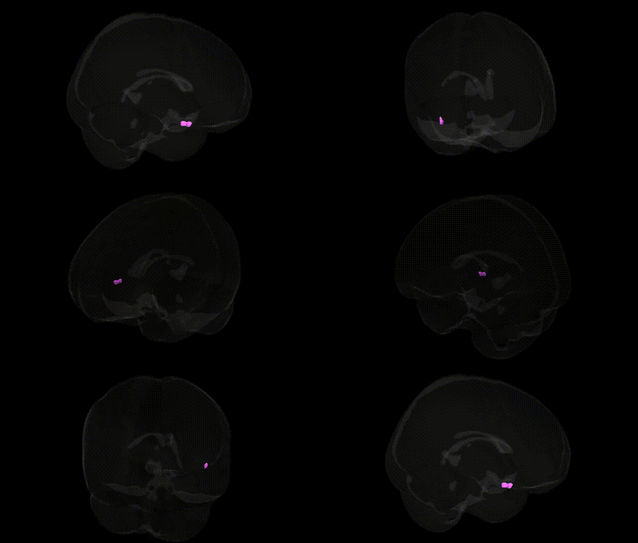
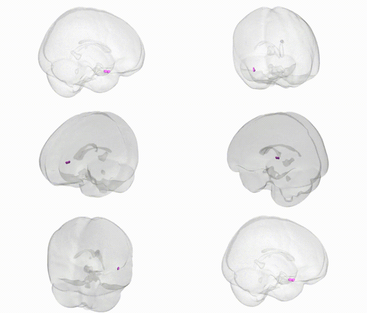
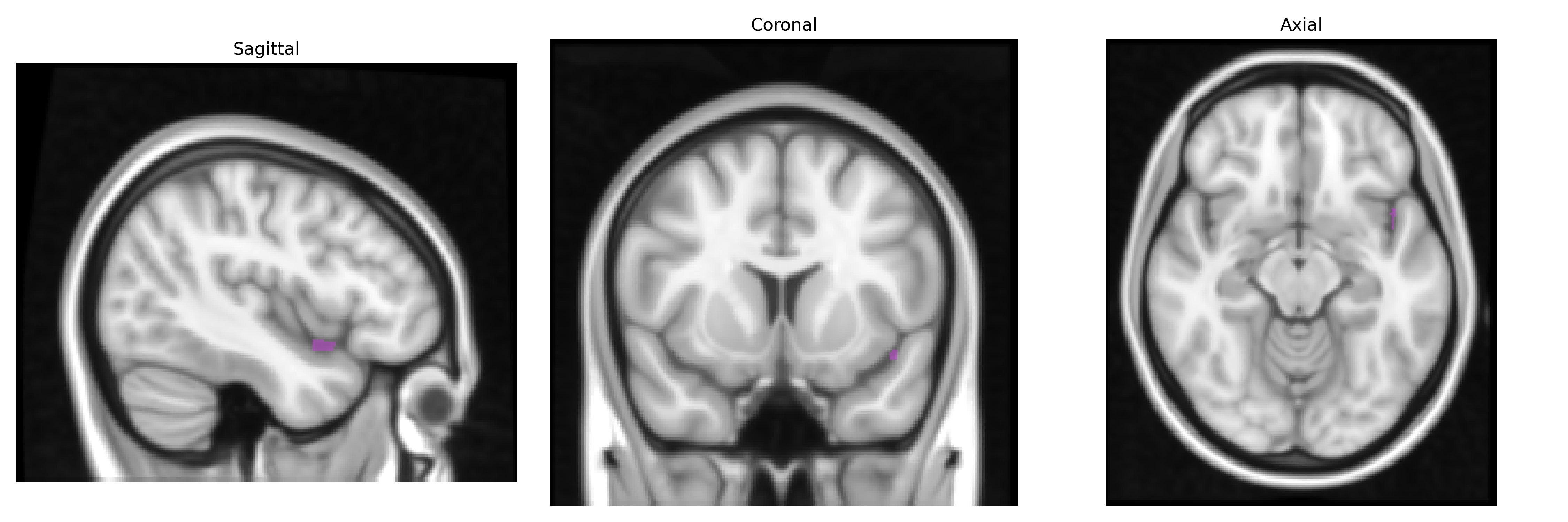
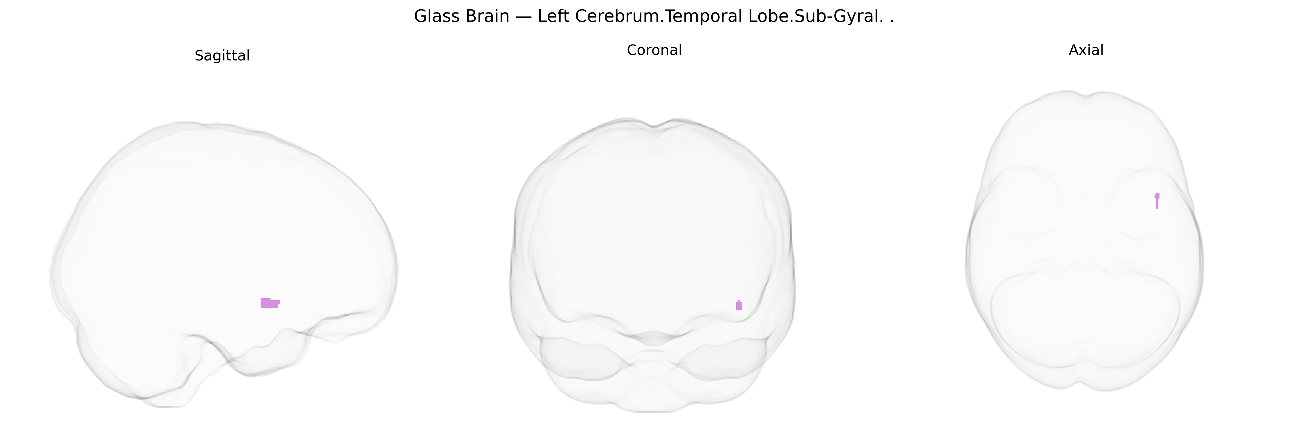
The bilateral Left Cerebrum.Temporal Lobe.Sub-Gyral region in the Talairach 1 mm atlas refers to subcortical white matter located beneath the cortical gyri of the left temporal lobe, including fibers that interconnect temporal cortical areas and link them with other lobes and subcortical structures. This region supports transmission of information between temporal cortical areas involved in auditory processing, language, semantic memory, and aspects of visual object and face recognition, as well as connections to limbic and frontal regions important for emotional processing and higher-order cognition. As a primarily descriptive anatomical label denoting deep white matter rather than a distinct gray-matter structure, it does not have a dedicated Wikipedia article; a related entry describing the overlying cortical territory is the Temporal lobe.
The bilateral left cerebrum temporal lobe sub-gyral region, corresponding roughly to deep portions of the superior, middle, and inferior temporal gyri in the Talairach 1 mm atlas, participates in language, semantic processing, auditory perception, and aspects of memory, and has been repeatedly implicated in imaging genetics and GWAS of brain structure and neuropsychiatric disease. Large-scale MRI–GWAS consortia (e.g., ENIGMA, UK Biobank) have identified numerous loci influencing temporal lobe cortical thickness, surface area, and volume, including common variants near or within genes involved in neurodevelopment, synaptic function, and axonal guidance such as HMGA2, MAPT, WNT and FGF pathway genes, and multiple loci on 3q, 6q, and 17q that modulate temporal cortical morphology. Left temporal sub-gyral regions show structural and functional alterations associated with risk variants for schizophrenia (e.g., in CACNA1C, MIR137, ZNF804A), bipolar disorder, major depression, and autism spectrum disorder, as well as in carriers of APOE ε4 and other Alzheimer’s disease–risk variants, where reduced temporal lobe volume and cortical thinning are characteristic phenotypes. GWAS of focal epilepsy have linked variants in genes such as SCN1A, GRIN2A, and others to temporal lobe epilepsy, which frequently shows volume loss and microstructural abnormalities in sub-gyral temporal cortex. Language-related genetic studies, including work on FOXP2 and polygenic scores for educational attainment and cognitive performance, have associated genetic variation with activation and structure in left temporal association cortex during speech and semantic tasks. Overall, the sub-gyral left temporal lobe emerges as a convergent site where polygenic influences on cortical development and connectivity intersect with risk architectures for psychiatric, neurodegenerative, epileptic, and cognitive traits, although few associations are specific to this exact Talairach-defined parcel, and most derive from broader temporal lobe or superior/middle temporal gyrus phenotypes.
Overview generated by GPT-4o (2026).
Region ID: 377
Hemisphere: bilateral
Atlas: Talairach labels 1mm
Full Quality Version: Download MP4
Full Quality Version: Download MP4
Wali Sidiqyar*, Gaurav Rudravaram*, Elyssa M. McMaster, Trent M. Schwartz, Adam M. Saunders, Kurt G. Schilling, Bennett A. Landman "Introducing SPINS: A Shared Public Visualization Library of Neuroanatomical Structures." Medical Imaging with Deep Learning- short paper
This resource is licensed under CC0 1.0 Universal (Public Domain).