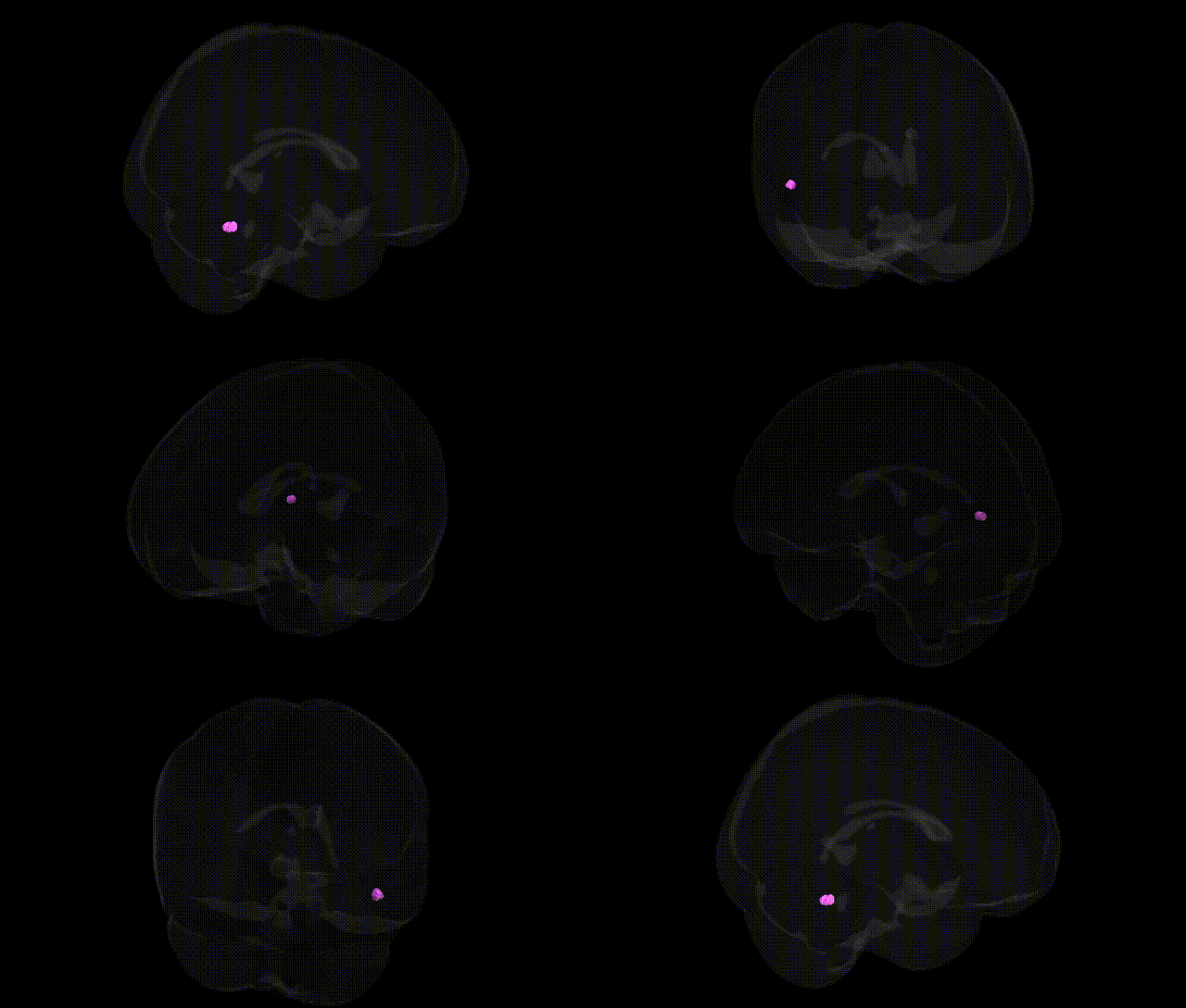
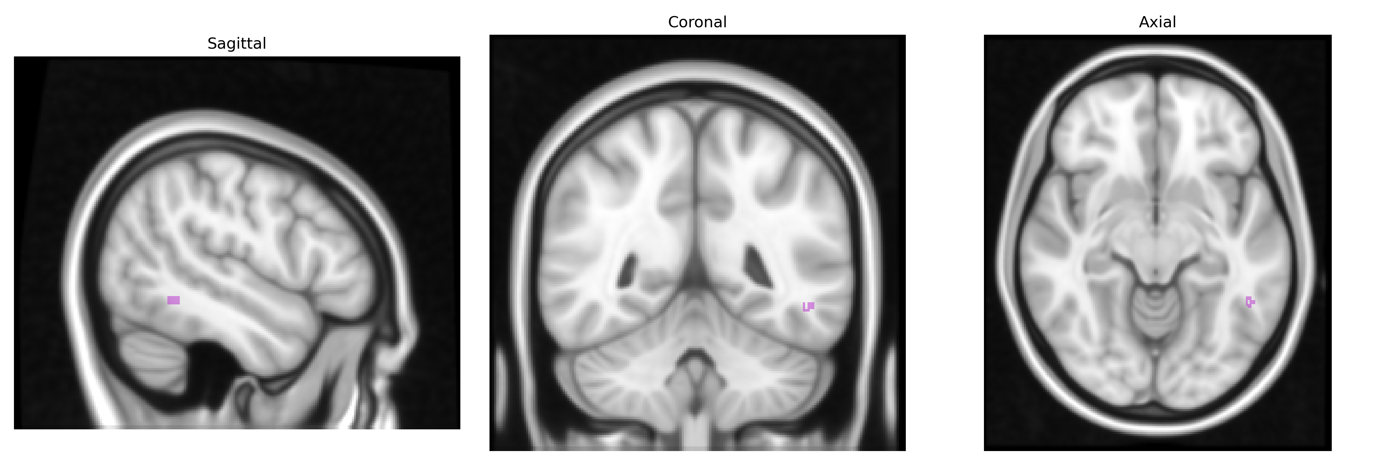
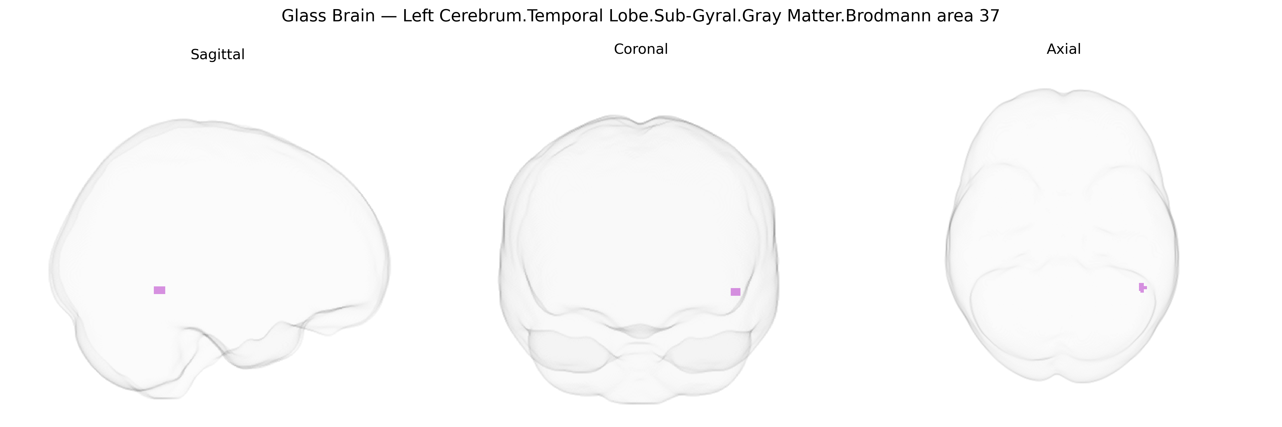
The bilateral Left Cerebrum.Temporal Lobe.Sub-Gyral.Gray Matter.Brodmann area 37 corresponds primarily to the left fusiform and inferior temporal gyri, a higher-order visual association cortex involved in complex object recognition, visual word processing, and particularly face and body perception. Situated in the ventral temporal lobe and extending into the occipitotemporal region, BA37 integrates inputs from earlier visual areas to support semantic processing of visually presented stimuli, linking visual features to stored knowledge about objects, words, and categories. Functionally, this region is crucial for multimodal integration of visual information with language and memory systems, and lesions can lead to deficits such as visual agnosia or impairments in reading and face recognition. There is no direct Wikipedia article for “Brodmann area 37” itself; a closely related and overlapping structure is the fusiform gyrus: Fusiform gyrus.
Brodmann area 37 in the bilateral left temporal sub-gyral gray matter, encompassing posterior inferior temporal and fusiform regions, has been repeatedly implicated in genetic studies of language, reading, and higher-order visual processing, with GWAS and imaging-genetics work linking variation in this region’s structure and function to several neurodevelopmental and psychiatric traits. Structural and functional measures in BA37/fusiform gyrus show heritability and associations with common variants near genes such as KIAA0319, DCDC2, and ROBO1 in dyslexia and reading-disorder cohorts, while FOXP2-related pathways and CNTNAP2 have been connected to language and speech phenotypes that engage this region during word and object recognition tasks. Large-scale imaging GWAS, including ENIGMA and UK Biobank analyses, have identified SNPs in or near genes such as HMGA2, IGF1, and MIRLET7BHG associated with temporal lobe cortical thickness and surface area, with effects often observed in inferior and fusiform temporal areas overlapping BA37. BA37 has also emerged in genetic risk mapping for autism spectrum disorder and schizophrenia, where polygenic risk scores and specific loci (for example near NRXN1, GRIN2A, and CRHR1 in some studies) are associated with altered fusiform/temporal activation or gray-matter volume during face processing, social cognition, or language tasks, and in Alzheimer’s disease and mild cognitive impairment, where variants in APOE (particularly ε4) and CLU, PICALM, and BIN1 correlate with atrophy or hypometabolism in lateral and inferior temporal cortex including BA37. Together, these findings indicate that BA37 is a key genetically modulated hub for visual word form and object recognition, language, and socio-cognitive processing, with its morphology and activation patterns influenced by polygenic architectures underlying reading disability, language impairment, ASD, schizophrenia, and neurodegenerative disease.
Overview generated by GPT-4o (2026).
Region ID: 332
Hemisphere: bilateral
Atlas: Talairach labels 1mm
Full Quality Version: Download MP4
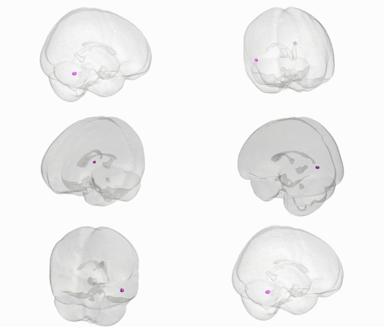
Full Quality Version: Download MP4
Wali Sidiqyar*, Gaurav Rudravaram*, Elyssa M. McMaster, Trent M. Schwartz, Adam M. Saunders, Kurt G. Schilling, Bennett A. Landman "Introducing SPINS: A Shared Public Visualization Library of Neuroanatomical Structures." Medical Imaging with Deep Learning- short paper
This resource is licensed under CC0 1.0 Universal (Public Domain).