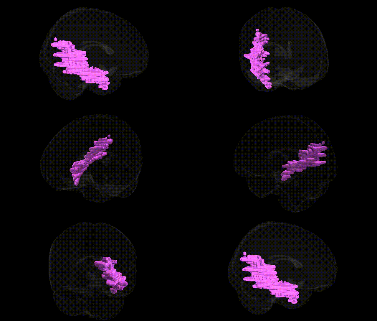
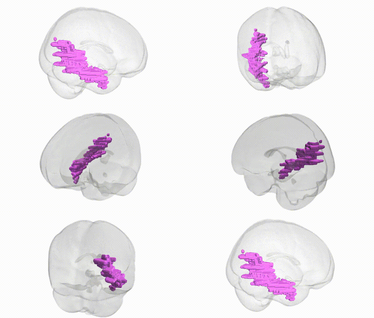
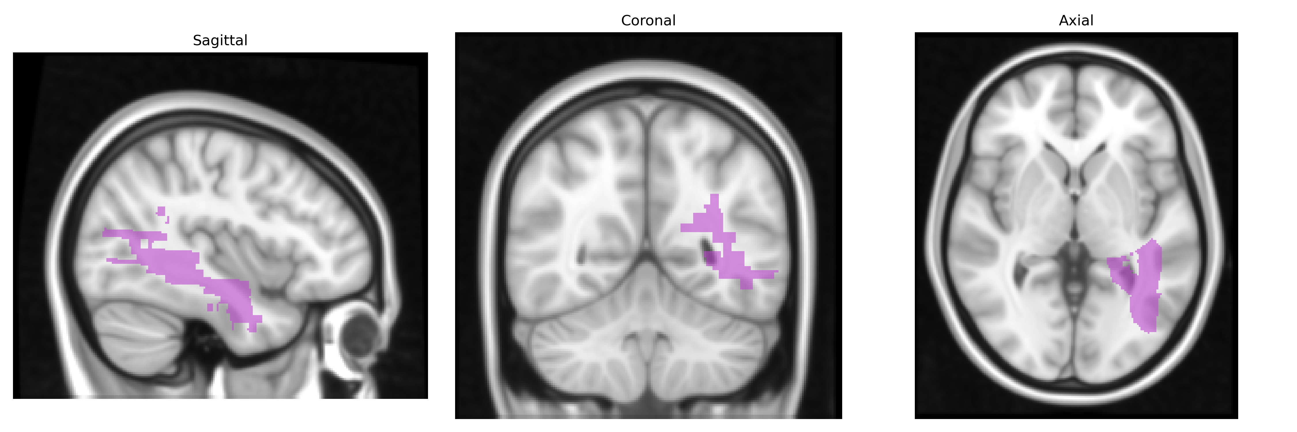
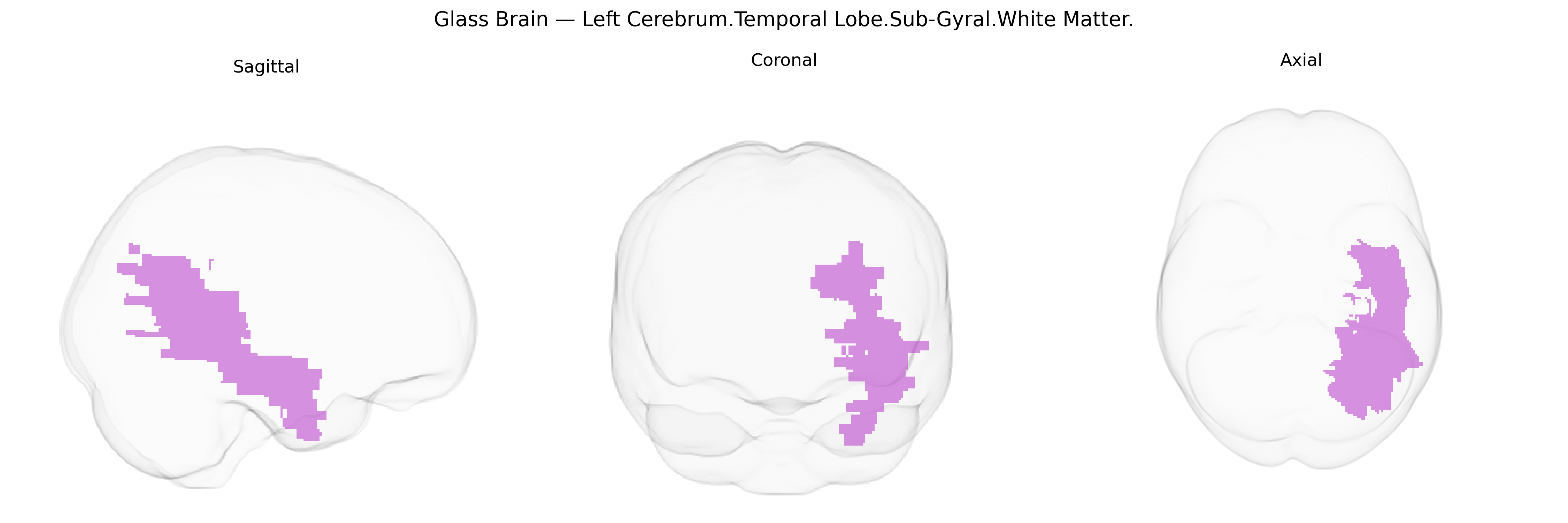
The bilateral Left Cerebrum.Temporal Lobe.Sub-Gyral.White Matter corresponds to deep white matter tracts located beneath the cortical gyri of the temporal lobe in the left cerebral hemisphere, extending medially and inferiorly to connect temporal cortical areas with frontal, parietal, limbic, and subcortical structures. These fibers include portions of association pathways such as the inferior longitudinal fasciculus and uncinate fasciculus, as well as projection fibers that relay auditory, language, memory, and multimodal sensory information to and from temporal cortical regions, including lateral temporal association cortex, medial temporal structures (such as the hippocampal formation), and perisylvian language areas. Functionally, this sub-gyral white matter supports higher-order auditory processing, semantic and phonological aspects of language, and aspects of episodic memory and object recognition by enabling rapid, coordinated communication between distributed temporal and extra-temporal networks. There is no direct Wikipedia article for this specific sub-gyral white matter region; a closely related structure is the Temporal lobe.
The bilateral left temporal sub-gyral white matter (Talairach: Left Cerebrum.Temporal Lobe.Sub-Gyral.White Matter) participates in long-range association pathways connecting temporal cortex with frontal, parietal, and limbic regions, and genetic influences on its microstructure and volume have been captured mainly through diffusion MRI and structural MRI GWAS. Twin and SNP-heritability studies show moderate heritability for temporal lobe white matter volume and fractional anisotropy, with large-scale consortia such as ENIGMA and UK Biobank implicating variants in genes involved in axonal guidance, myelination, and neurodevelopment (e.g., NRG1/NRG3, CNTNAP2, MAG, PLP1, and loci near LINGO1 and ROBO1) in inter-individual variation of temporal white matter integrity. Polygenic risk for schizophrenia, bipolar disorder, and major depression is associated with reduced integrity of left temporal white matter tracts, and risk loci in or near CACNA1C, ZNF804A, and DISC1 have been linked to altered temporal connectivity, including regions overlapping sub-gyral white matter. GWAS of language, reading, and educational attainment identify variants (e.g., in FOXP2-regulated networks and DCDC2/KIAA0319 clusters) associated with temporal white matter pathways supporting phonological and semantic processing, while autism spectrum disorder and specific language impairment risk genes (e.g., CNTNAP2, NRXN1) have been tied to atypical temporal white matter organization. Neurodegenerative and demyelinating disease genetics (e.g., APOE ε4 in Alzheimer’s disease, HLA-DRB1*15:01 and other immune-related loci in multiple sclerosis) show convergent effects on temporal white matter atrophy and lesion burden, suggesting that this region is a common anatomical substrate where diverse genetic risks for psychiatric, cognitive, and neurodegenerative phenotypes manifest through alterations of white matter structure and connectivity.
Overview generated by GPT-4o (2026).
Region ID: 51
Hemisphere: bilateral
Atlas: Talairach labels 1mm
Full Quality Version: Download MP4
Full Quality Version: Download MP4
Wali Sidiqyar*, Gaurav Rudravaram*, Elyssa M. McMaster, Trent M. Schwartz, Adam M. Saunders, Kurt G. Schilling, Bennett A. Landman "Introducing SPINS: A Shared Public Visualization Library of Neuroanatomical Structures." Medical Imaging with Deep Learning- short paper
This resource is licensed under CC0 1.0 Universal (Public Domain).