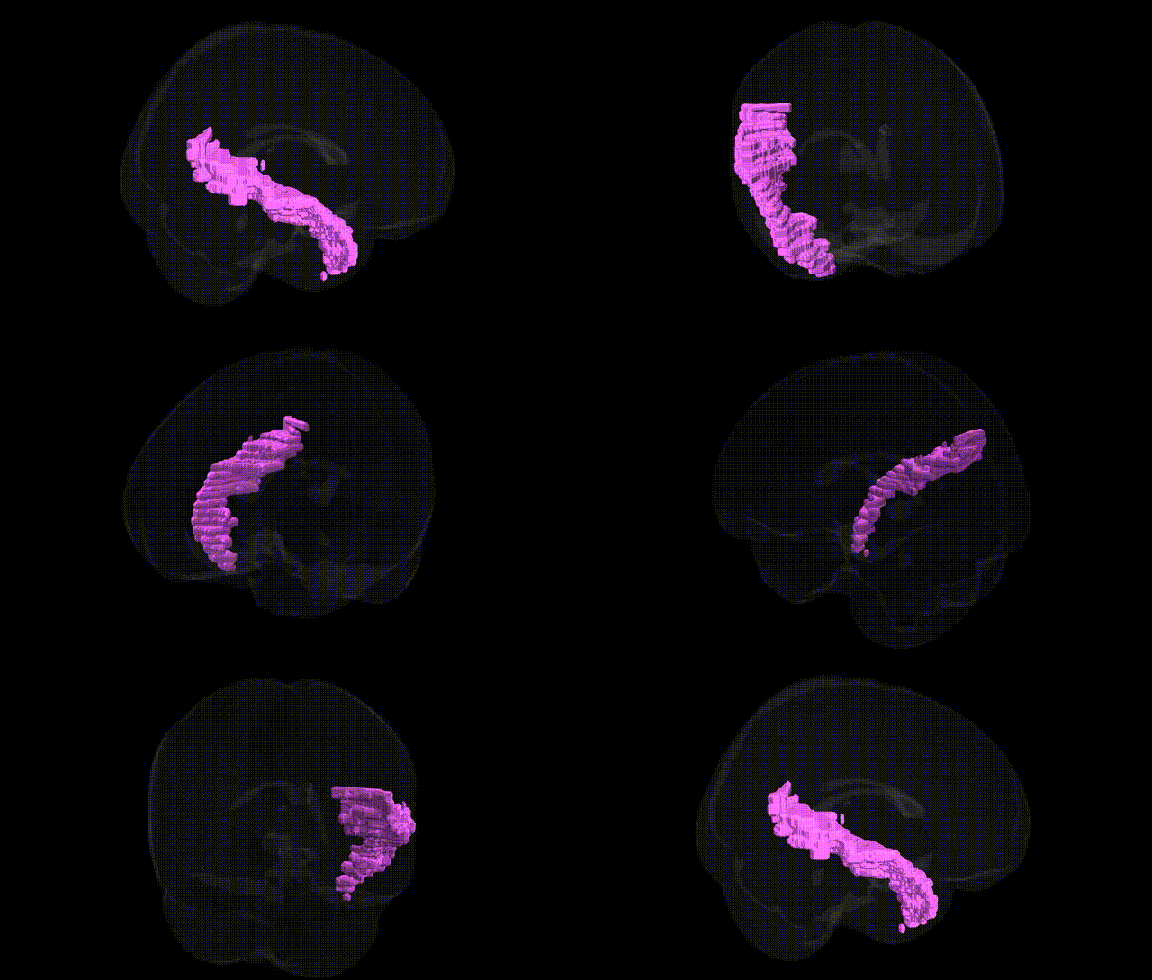
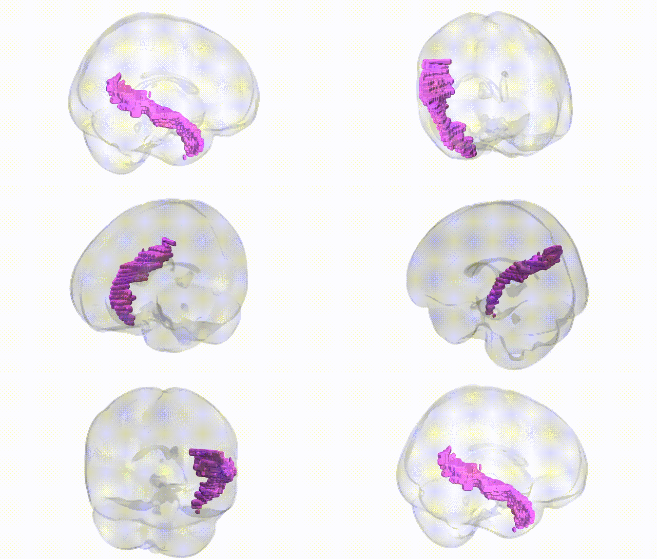
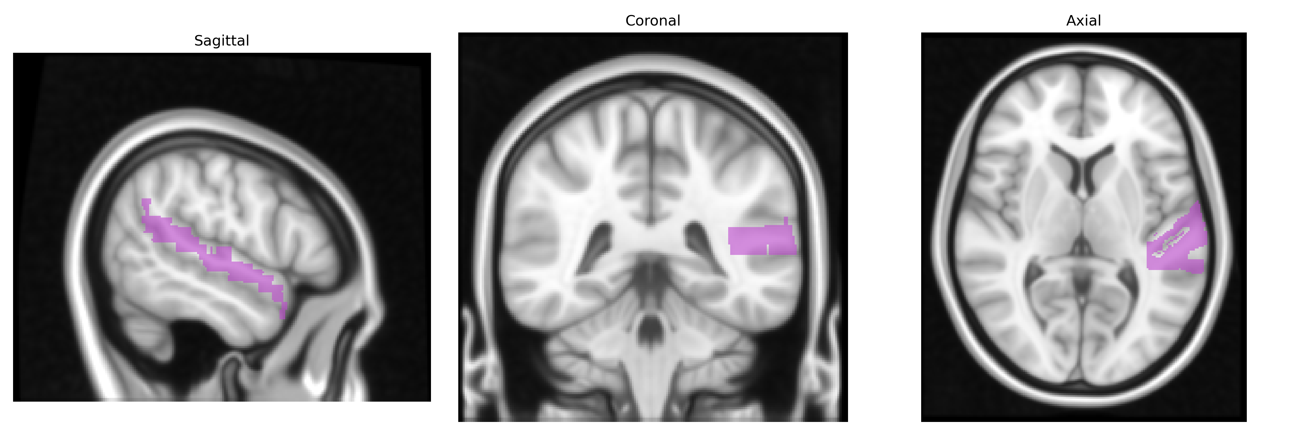
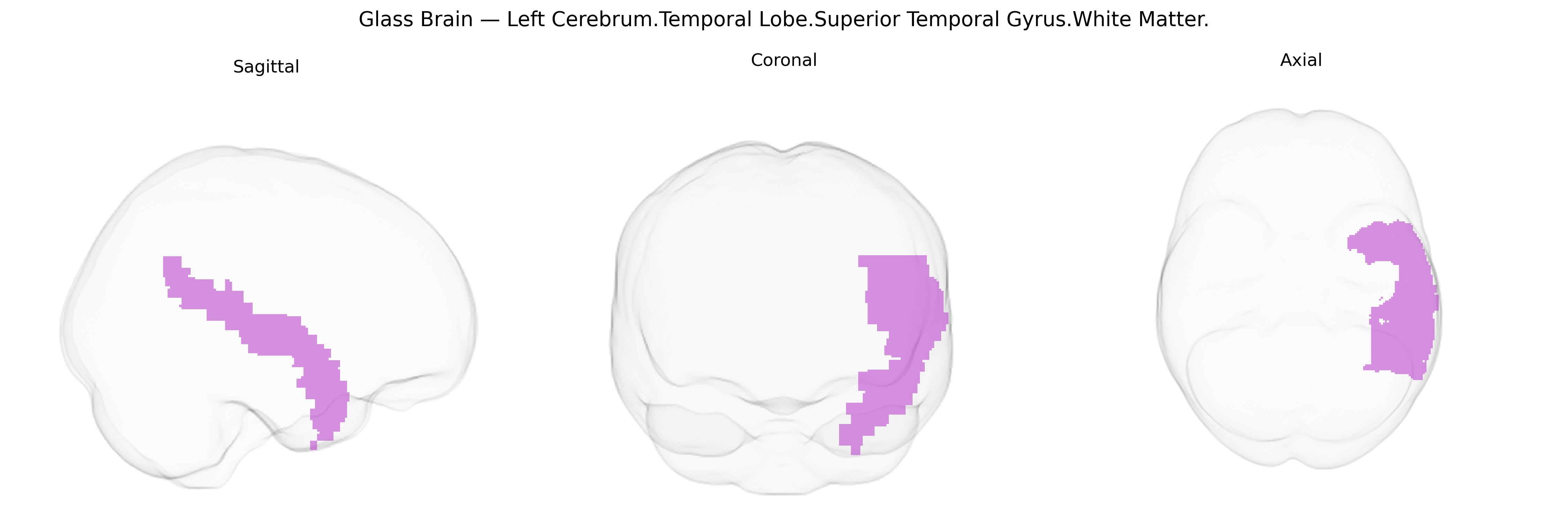
The bilateral Left Cerebrum.Temporal Lobe.Superior Temporal Gyrus.White Matter corresponds to the deep myelinated fiber pathways underlying the superior temporal gyrus in the left temporal lobe, including association and projection fibers that interconnect primary and secondary auditory cortices, language-related regions (such as Wernicke’s area), and other temporal, parietal, and frontal areas. These white matter tracts support rapid transmission of auditory, phonological, and higher-order linguistic information, as well as integration of auditory input with multimodal sensory and cognitive networks. They form part of larger temporal lobe white matter systems, including segments of the arcuate fasciculus and superior longitudinal fasciculus, thereby contributing critically to speech perception, language comprehension, and aspects of auditory memory and social communication processing. Related cortical information can be found at Superior temporal gyrus.
The bilateral Left Cerebrum.Temporal Lobe.Superior Temporal Gyrus.White Matter, corresponding to major association fibers linking auditory, language, and multimodal integration cortices, is implicated in several genetically influenced traits and disorders, though GWAS typically map to broader superior temporal or temporal-lobe white-matter measures rather than this exact Talairach label. Twin and family studies show high heritability of superior temporal gyrus (STG) thickness, volume, and white-matter microstructure, with common variants in genes regulating neurodevelopment, myelination, and synaptic function (e.g., NRG1/ERBB signaling, CNTNAP2, DISC1, and FOXP2-related pathways) repeatedly associated with temporal-lobe structure and connectivity. Large imaging–genetics consortia such as ENIGMA and UK Biobank report genome-wide significant loci for temporal lobe and STG surface area and white-matter fractional anisotropy, including variants near genes involved in axon guidance and oligodendrocyte biology (e.g., DCC, ROBO family, and myelin-related genes), and polygenic scores for global brain morphology partially explain variance in superior temporal white-matter integrity. Clinically, genetic risk for schizophrenia, bipolar disorder, and major depression shows robust associations with reduced STG volume and altered temporal white-matter tracts, with psychosis risk loci (e.g., CACNA1C, GRIN2A, complement pathway genes such as C4) linked to temporal-lobe structural differences in ENIGMA and other GWAS-derived imaging studies. Autism spectrum disorder and language-related phenotypes show convergent evidence implicating superior temporal regions, where rare and common variants in synaptic and neurodevelopmental genes (e.g., SHANK3, NRXN1, 16p11.2 CNVs, FOXP2 network genes) are associated with atypical superior temporal cortical and white-matter development, especially in language and social-communication networks. Additionally, GWAS of reading, educational attainment, and verbal ability identify polygenic influences on temporal-lobe anatomy and connectivity, suggesting that genetic architectures underlying cognitive and psychiatric traits partly exert effects through structural variation in superior temporal white matter, although current evidence remains indirect and regionally coarse rather than specific to this precise Talairach-defined parcel.
Overview generated by GPT-4o (2026).
Region ID: 39
Hemisphere: bilateral
Atlas: Talairach labels 1mm
Full Quality Version: Download MP4
Full Quality Version: Download MP4
Wali Sidiqyar*, Gaurav Rudravaram*, Elyssa M. McMaster, Trent M. Schwartz, Adam M. Saunders, Kurt G. Schilling, Bennett A. Landman "Introducing SPINS: A Shared Public Visualization Library of Neuroanatomical Structures." Medical Imaging with Deep Learning- short paper
This resource is licensed under CC0 1.0 Universal (Public Domain).