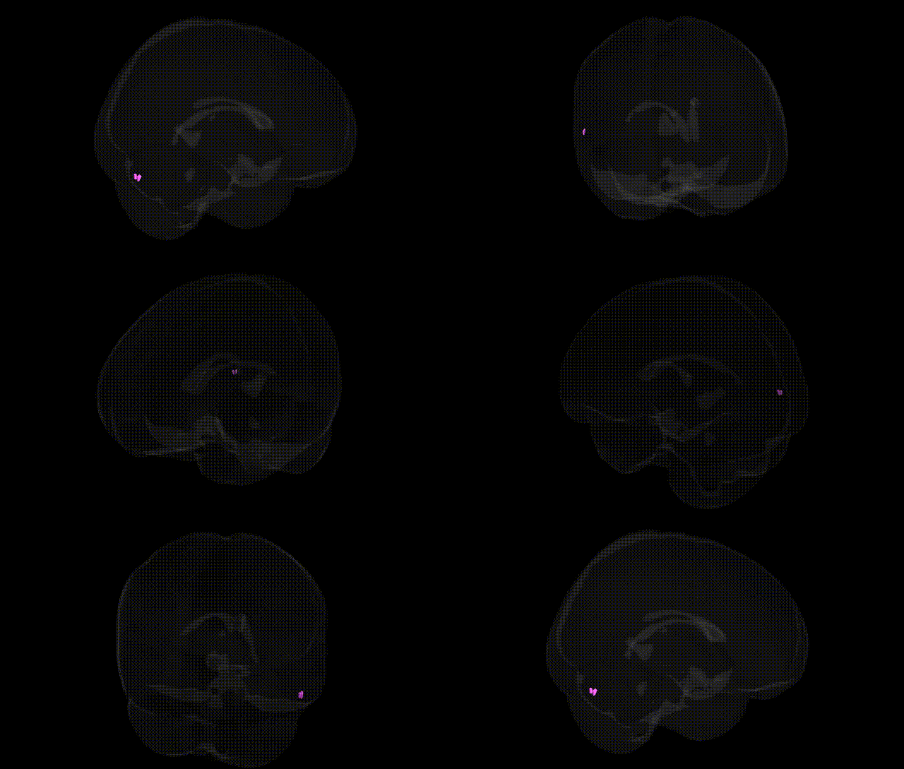
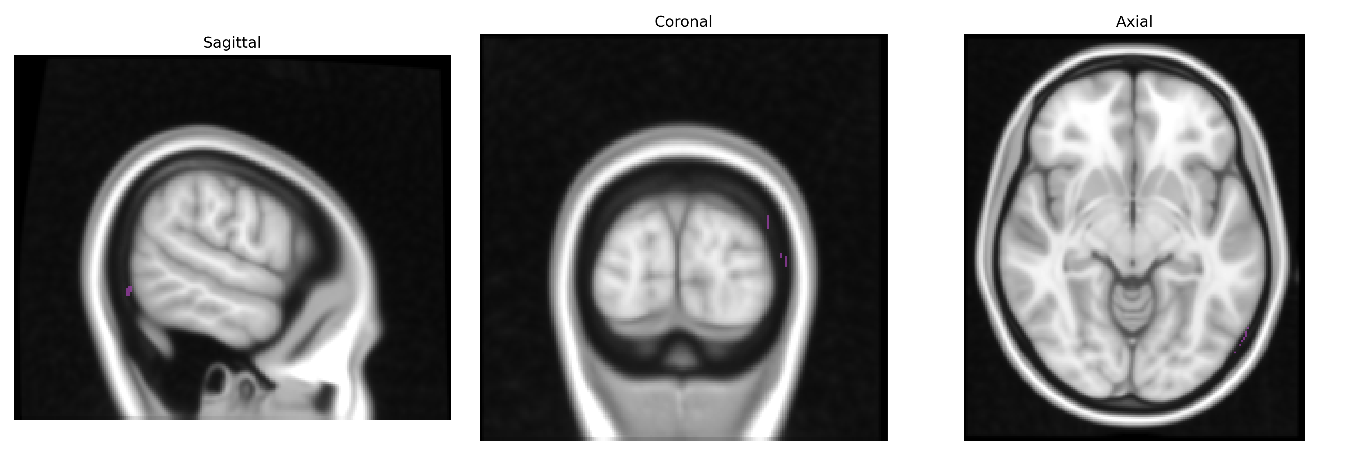
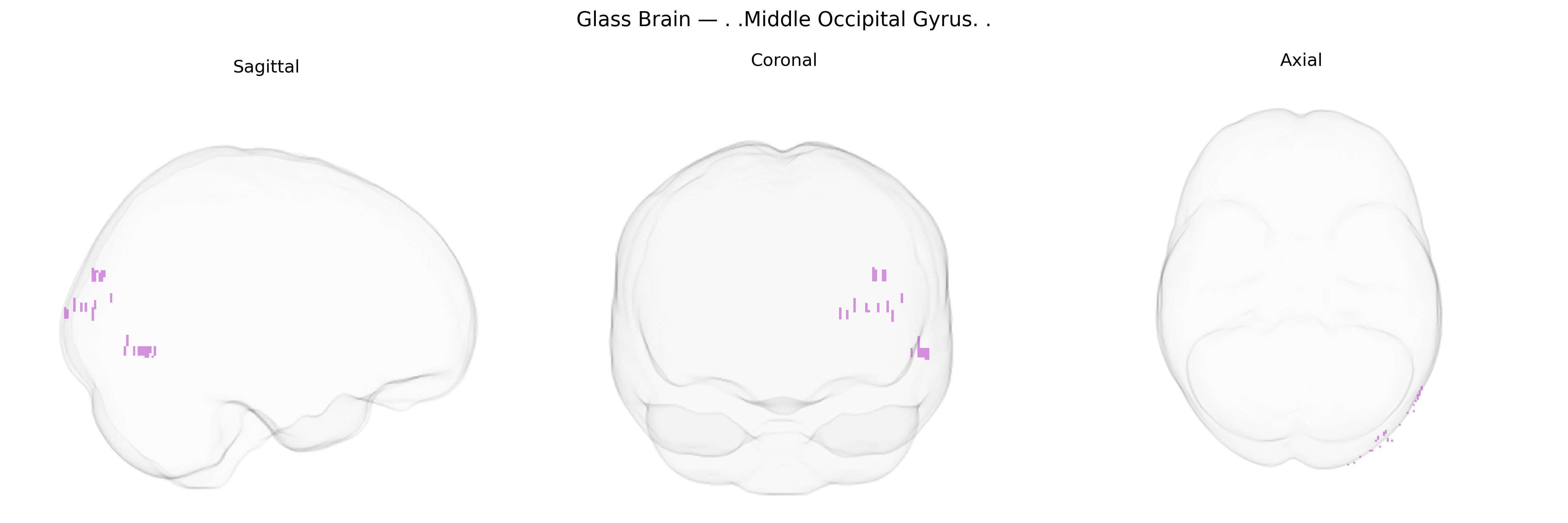
The bilateral Middle Occipital Gyrus is a cortical region located in the middle portion of the occipital lobe of both hemispheres, lying between the superior and inferior occipital gyri and extending along the lateral surface of the posterior brain. It is primarily associated with intermediate-level visual processing, including analysis of form, motion, and spatial relationships, and contributes to integration of visual information for perception and visually guided behavior. This gyrus participates in dorsal and ventral visual stream interactions, linking early visual areas with higher-order associative cortices involved in object recognition and visuospatial attention. There is no direct link for the Middle Occipital Gyrus; see the related structure Occipital lobe.
The bilateral middle occipital gyrus, a core visual association region, has been implicated in several genetic and GWAS-based findings primarily through imaging genetics and neuropsychiatric studies rather than locus-specific investigations of the Talairach 1 mm atlas label itself. Variants in genes involved in synaptic plasticity and neurodevelopment—such as BDNF (e.g., Val66Met), COMT, and DISC1—have been repeatedly associated with altered occipital activation or cortical thickness during visual processing and cognitive tasks, while large-scale imaging GWAS (e.g., ENIGMA, UK Biobank) have identified polygenic influences on occipital lobe surface area and volume, often enriched for neurodevelopmental and axon guidance pathways. Occipital structural or functional alterations linked to genetic risk scores have been reported in schizophrenia, major depressive disorder, autism spectrum disorder, and ADHD, suggesting that common risk variants for these conditions partly exert effects through visual association cortices including the middle occipital gyrus. Additionally, GWAS of traits such as general cognitive ability, educational attainment, and reading/visual word recognition have shown associations with occipital and temporo-occipital measures, indicating that polygenic factors influencing higher cognitive and perceptual skills also modulate morphology and activity in this region. While no single gene is uniquely or specifically tied to the Talairach-defined bilateral middle occipital gyrus, convergent evidence from neuroimaging genetics supports a role for broadly distributed neurodevelopmental and synaptic genes in shaping its structure and function, often in the context of psychiatric, cognitive, and sensory processing phenotypes.
Overview generated by GPT-4o (2026).
Region ID: 317
Hemisphere: bilateral
Atlas: Talairach labels 1mm
Full Quality Version: Download MP4
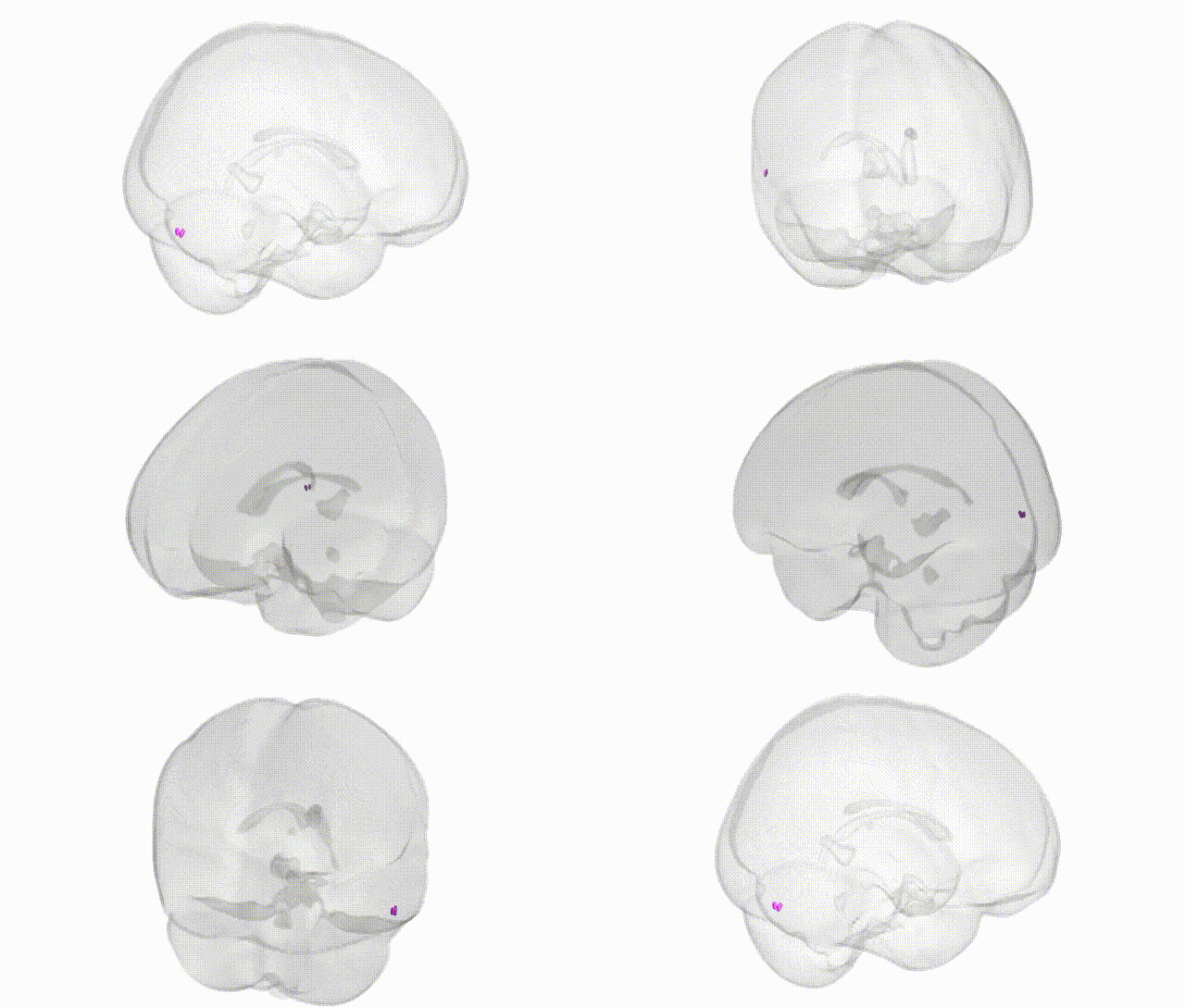
Full Quality Version: Download MP4
Wali Sidiqyar*, Gaurav Rudravaram*, Elyssa M. McMaster, Trent M. Schwartz, Adam M. Saunders, Kurt G. Schilling, Bennett A. Landman "Introducing SPINS: A Shared Public Visualization Library of Neuroanatomical Structures." Medical Imaging with Deep Learning- short paper
This resource is licensed under CC0 1.0 Universal (Public Domain).