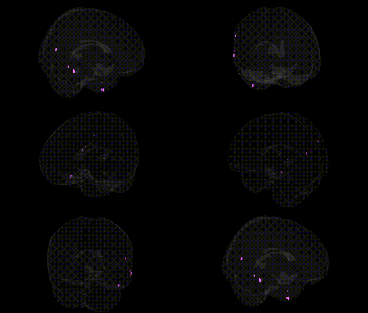
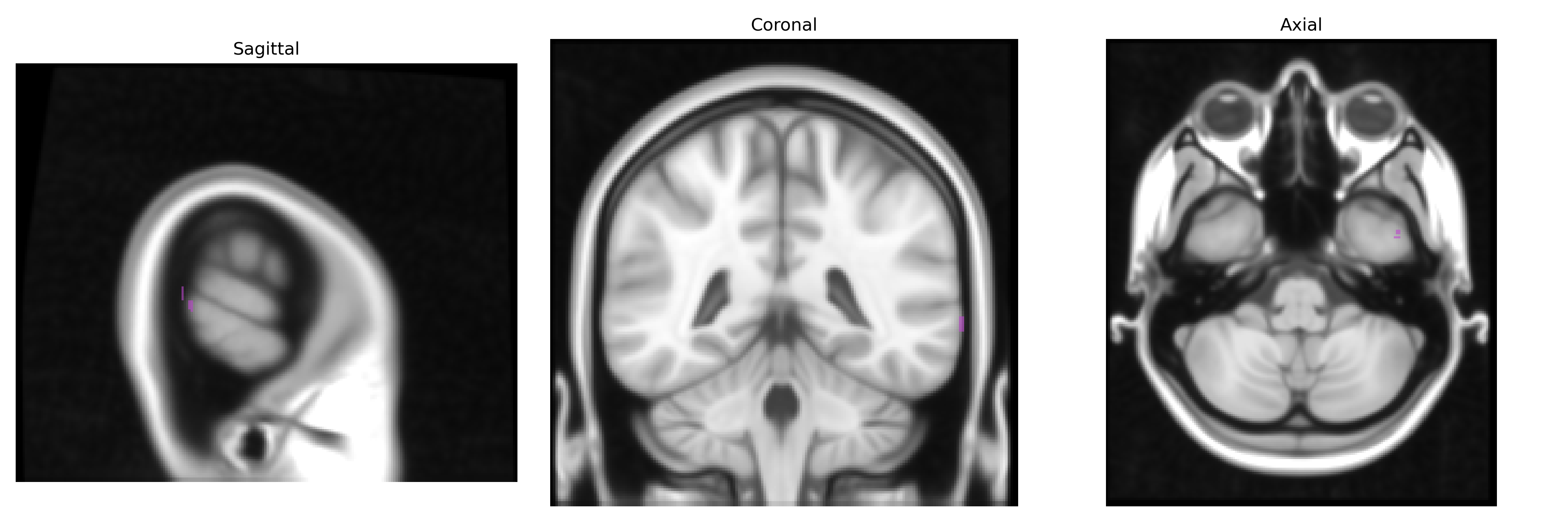
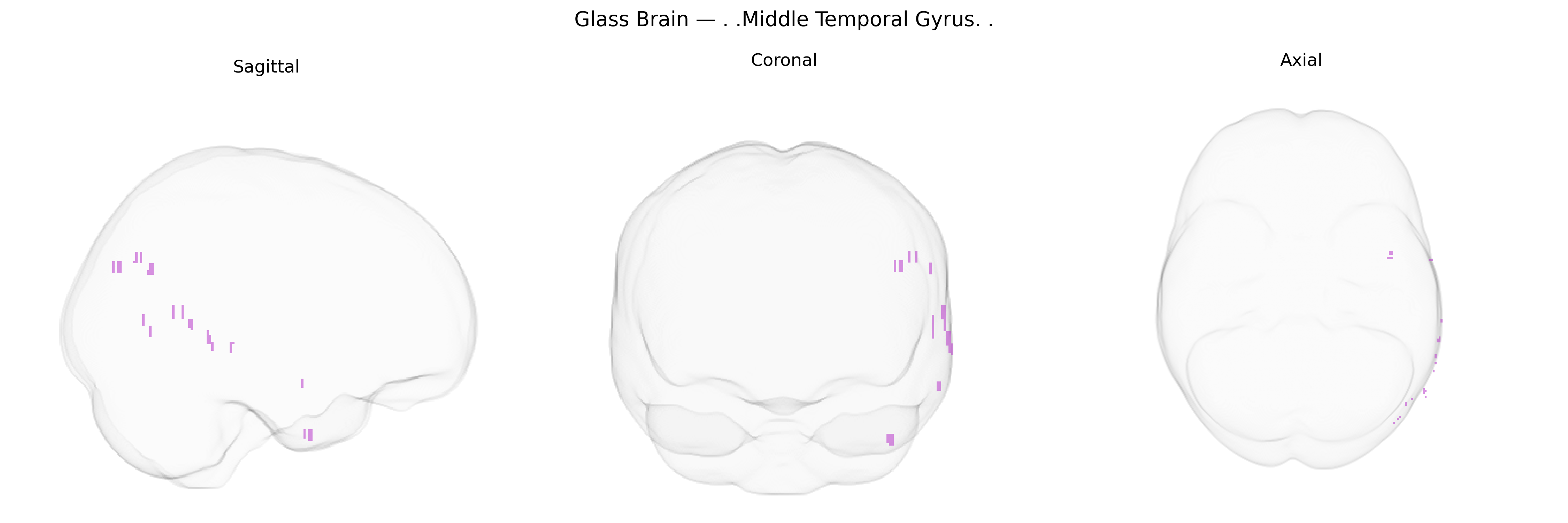
The bilateral Middle Temporal Gyrus is a cortical region of the lateral temporal lobe, situated between the superior and inferior temporal gyri, extending from the temporal pole anteriorly toward the angular gyrus posteriorly. It is involved in multimodal association processing, including semantic memory, language comprehension, higher-order visual perception (such as motion and biological form), and social-cognitive functions like theory of mind. Cytoarchitectonically, it encompasses portions of Brodmann areas 21 and 22, with connectivity to frontal language regions, inferior parietal cortex, and limbic structures via major white matter pathways such as the superior longitudinal fasciculus and uncinate fasciculus. Functional imaging frequently implicates this region in auditory and visual semantic processing, narrative comprehension, and integration of contextual information across modalities. Middle temporal gyrus
The bilateral Middle Temporal Gyrus (MTG), as defined in the Talairach 1 mm atlas, has been repeatedly implicated in genetic studies of language, social cognition, and psychiatric and neurodevelopmental disorders. GWAS and imaging-genetics work indicate that common variants influencing cortical thickness and surface area in temporal association cortices, including MTG, cluster in genes related to synaptic development and neuronal signaling (for example, variants near genes such as KIAA0319 and DCDC2 implicated in dyslexia, and CNTNAP2 and FOXP2 associated with language and communication phenotypes), with MTG often emerging as a key locus for phonological and semantic processing. Large consortia (e.g., ENIGMA, UK Biobank) have shown that polygenic risk scores for schizophrenia, autism spectrum disorder, and major depressive disorder are associated with structural and functional alterations in bilateral MTG, linking risk variants in glutamatergic, GABAergic, and synaptic plasticity pathways to temporal lobe morphology and connectivity. In social and cognitive GWAS, MTG activation and volume correlate with variants associated with social communication ability, theory of mind, and educational attainment, while Alzheimer’s disease and frontotemporal dementia risk loci (such as APOE and MAPT region variants) are associated with atrophy patterns that prominently involve temporal association cortex, including MTG. Collectively, genetic evidence supports the MTG as a convergence zone where variants affecting language, social cognition, and psychiatric vulnerability exert measurable effects on brain structure and function.
Overview generated by GPT-4o (2026).
Region ID: 54
Hemisphere: bilateral
Atlas: Talairach labels 1mm
Full Quality Version: Download MP4
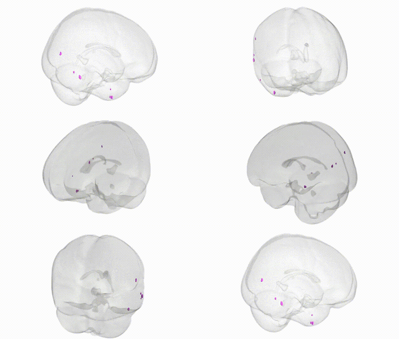
Full Quality Version: Download MP4
Wali Sidiqyar*, Gaurav Rudravaram*, Elyssa M. McMaster, Trent M. Schwartz, Adam M. Saunders, Kurt G. Schilling, Bennett A. Landman "Introducing SPINS: A Shared Public Visualization Library of Neuroanatomical Structures." Medical Imaging with Deep Learning- short paper
This resource is licensed under CC0 1.0 Universal (Public Domain).