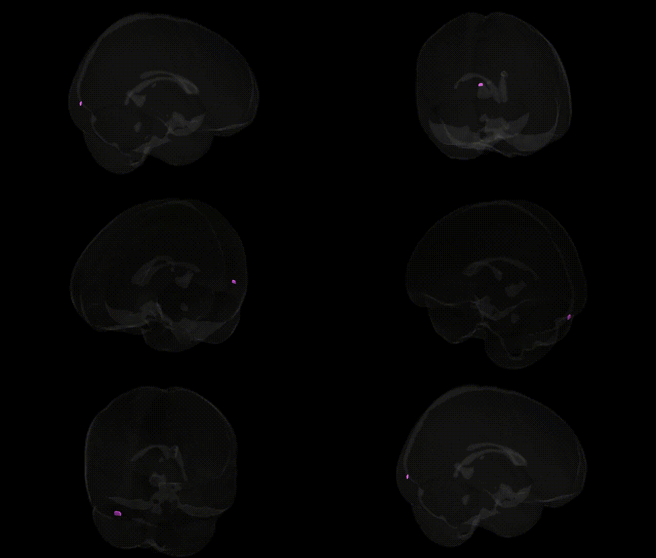
The bilateral occipital lobe lingual gyrus is a ventromedial occipital cortical region that extends along the medial aspect of the occipital lobe into the posterior temporal region, forming part of the inferior bank of the calcarine sulcus. It is primarily involved in visual processing, including analysis of complex visual patterns, letter and word recognition, and aspects of scene and color processing, and is a key component of the ventral visual (“what”) pathway. Functionally, it participates in transforming early visual input into higher-order representations, contributing to reading, visual memory, and object identification, and shows strong connectivity with other occipital, temporal, and parietal association areas. Clinically, lesions in this region can contribute to visual field defects and higher-order visual disturbances, such as alexia without agraphia when involvement extends into adjacent structures like the fusiform and splenial regions. Lingual gyrus
The bilateral occipital lobe lingual gyrus, as defined in the Talairach 1 mm atlas, has been implicated in several genetic and GWAS-based associations primarily through imaging–genetics and neuropsychiatric studies, although findings are often reported under broader occipital/ventral visual regions rather than this label alone. Variants in genes involved in synaptic function, neurodevelopment, and myelination (such as BDNF, NRG1, and CNTNAP2) have been associated with altered occipital and lingual gyrus structure or activation in tasks involving visual processing, reading, and face/word recognition, with some studies linking BDNF Val66Met and dyslexia-related loci to lingual/ventral occipitotemporal activation differences. Large-scale GWAS of cortical thickness and surface area (e.g., ENIGMA and UK Biobank consortia) have identified loci near genes such as MIR924HG, DAAM1, and others associated with occipital cortical metrics that encompass the lingual gyrus, with effects tied to pathways in axon guidance and neuronal differentiation. Lingual gyrus morphometry and function show heritable components and have been linked genetically to neurodevelopmental and psychiatric conditions, including autism spectrum disorder, schizophrenia, and major depressive disorder, where risk loci (for example in CACNA1C, ZNF804A, and other polygenic load) correlate with altered activation or volume in this region during visual, emotional, or memory tasks. Additionally, GWAS of migraine, visual hallucinations in Parkinson’s disease, and occipital lobe epilepsy have implicated variants in ion channel and neurotransmission genes (e.g., CACNA1A and GABA-related genes) that modulate excitability in occipital visual cortex, including the lingual gyrus, while polygenic risk for Alzheimer’s disease and small vessel disease shows associations with occipital/lingual atrophy or hypometabolism patterns, linking this region indirectly to APOE and other dementia-related loci.
Overview generated by GPT-4o (2026).
Region ID: 241
Hemisphere: bilateral
Atlas: Talairach labels 1mm
Full Quality Version: Download MP4
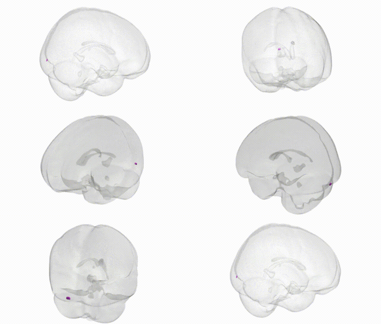
Full Quality Version: Download MP4
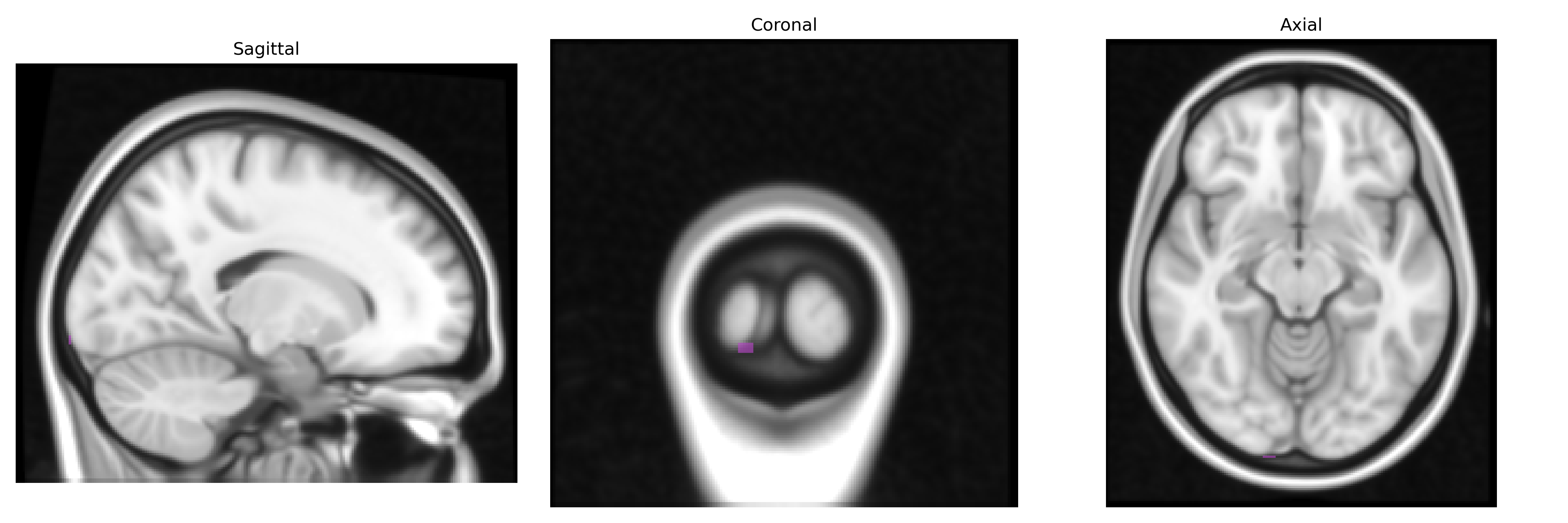
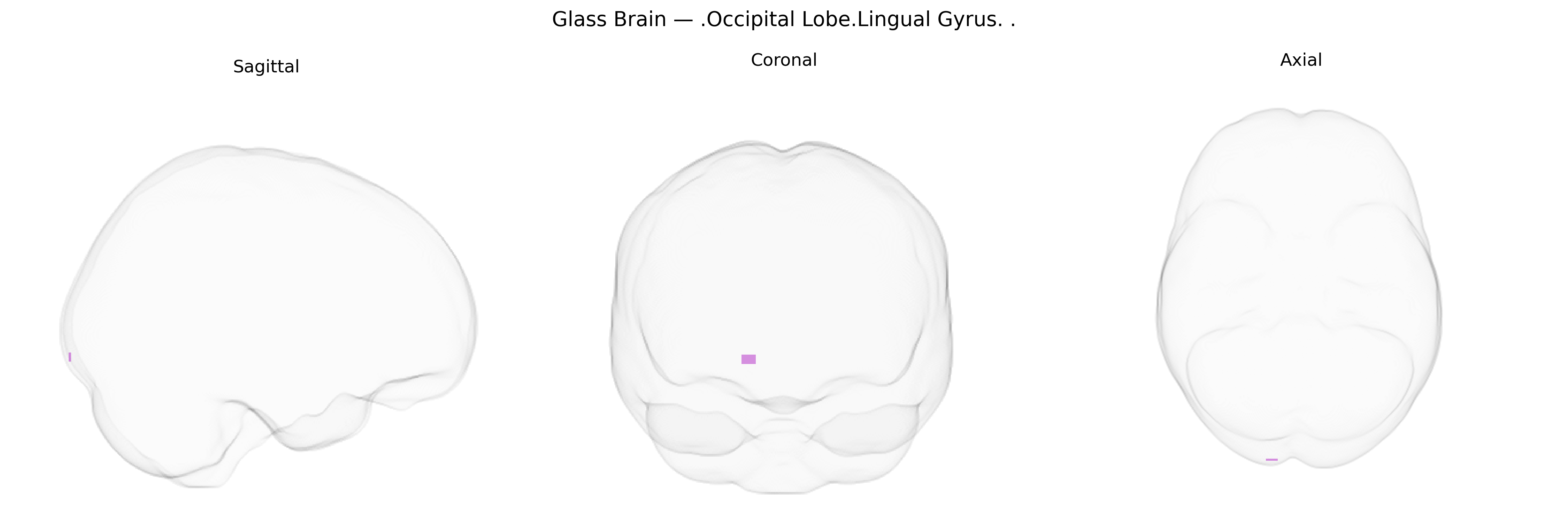
Wali Sidiqyar*, Gaurav Rudravaram*, Elyssa M. McMaster, Trent M. Schwartz, Adam M. Saunders, Kurt G. Schilling, Bennett A. Landman "Introducing SPINS: A Shared Public Visualization Library of Neuroanatomical Structures." Medical Imaging with Deep Learning- short paper
This resource is licensed under CC0 1.0 Universal (Public Domain).