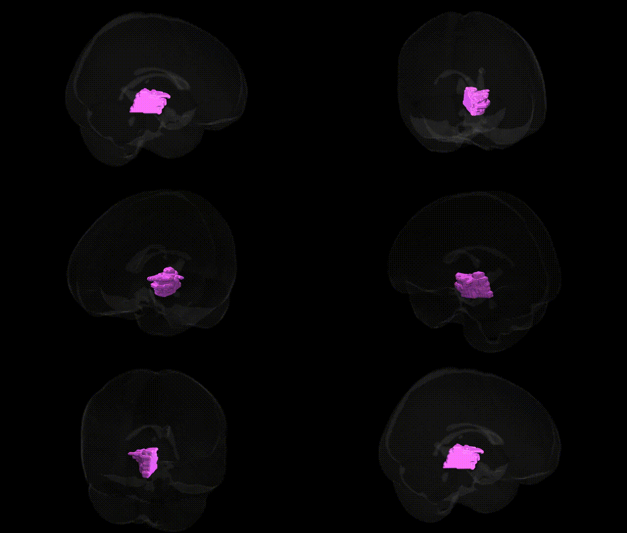
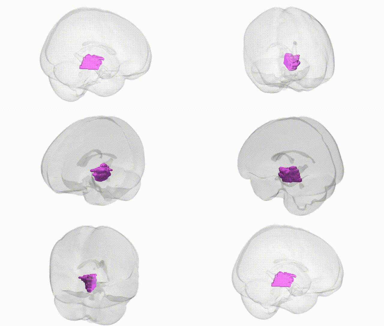
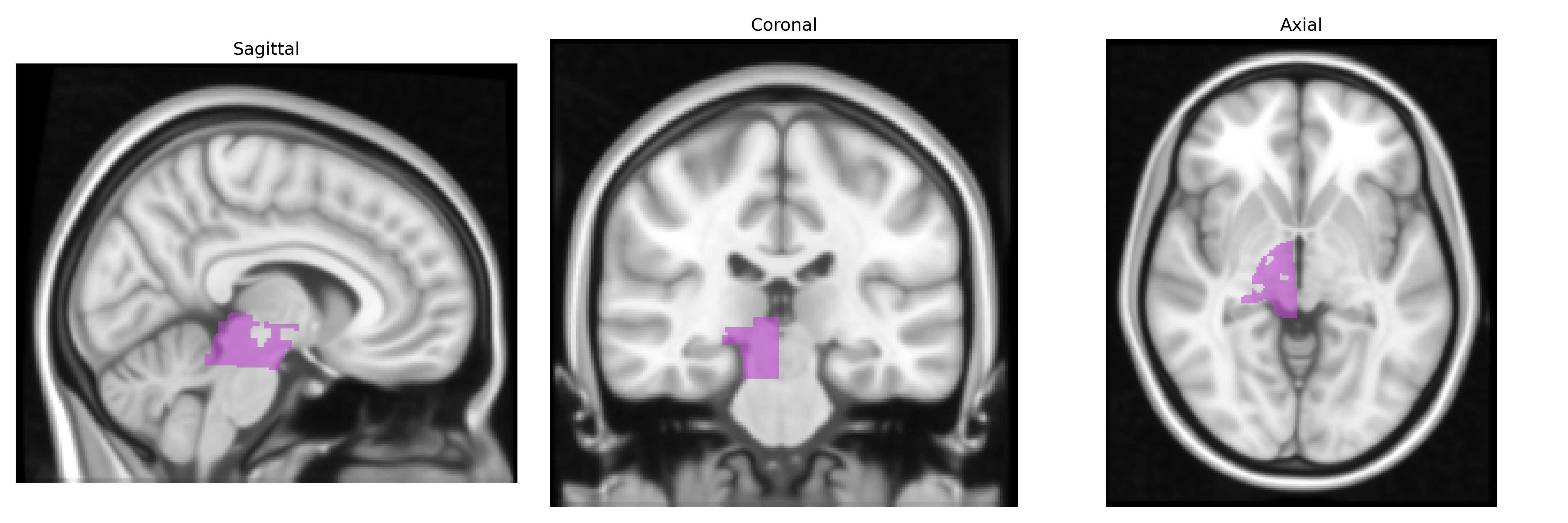
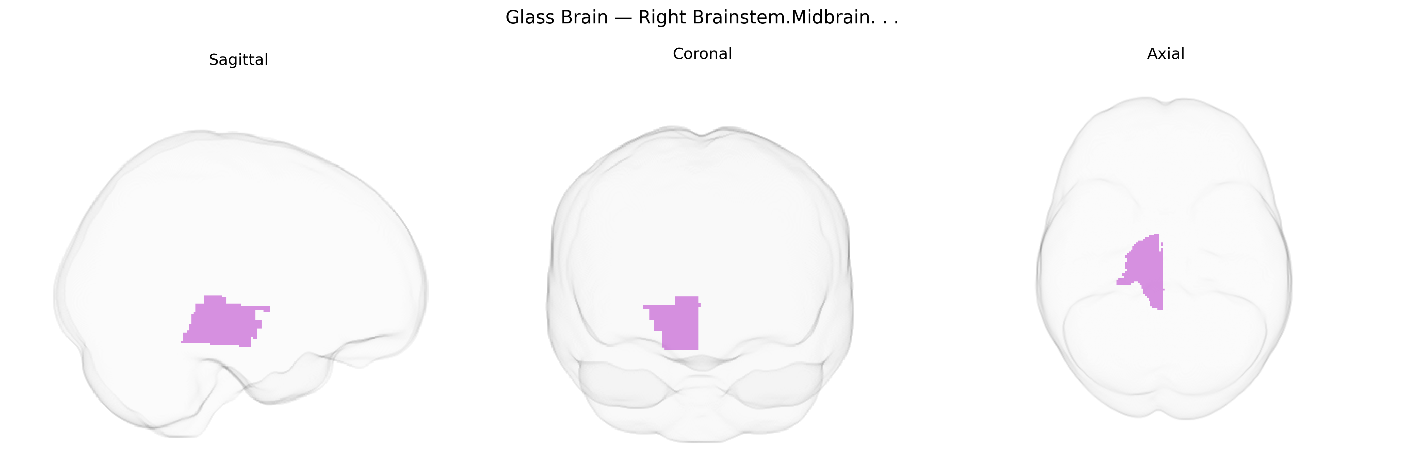
The bilateral Right Brainstem.Midbrain region in the Talairach 1 mm atlas corresponds to the right-sided portion of the midbrain, a central segment of the brainstem located between the pons and the diencephalon. This area contains key structures such as the tectum (including the superior and inferior colliculi), tegmentum, and cerebral peduncles, which collectively participate in motor control, auditory and visual reflex pathways, arousal, and the modulation of pain and autonomic functions. Major ascending and descending fiber tracts traverse this region, including components of the corticospinal, corticobulbar, and sensory pathways, making it a crucial conduit between the forebrain and lower brainstem/spinal cord. Functionally, the right midbrain contributes to the integration of sensorimotor information, coordination of eye movements and head-orienting responses, and regulation of consciousness and wakefulness through connections with the reticular formation and thalamus.
The bilateral right brainstem/midbrain region, as defined in the Talairach 1 mm atlas, is functionally and genetically linked to multiple neuropsychiatric and neurodegenerative conditions, although GWAS typically implicate genes and networks rather than this anatomical label directly. Genetic studies of midbrain dopaminergic systems identify variants in genes such as DRD2, DRD3, DAT1/SLC6A3, and COMT that influence midbrain dopamine signaling and have been associated with schizophrenia, bipolar disorder, attention-deficit/hyperactivity disorder, substance use disorders, and cognitive traits like working memory and executive function. GWAS of Parkinson’s disease and related movement disorders repeatedly implicate loci including SNCA, LRRK2, GBA, MAPT, and VPS35, which affect the integrity of midbrain structures, particularly the substantia nigra, contributing to motor symptoms and dopaminergic cell loss. Variants in TCF4, CACNA1C, and other synaptic and calcium-channel genes have been associated with brainstem/midbrain activation and connectivity patterns in imaging genetics studies of anxiety, depression, and psychosis. Additionally, large-scale imaging GWAS (e.g., ENIGMA and UK Biobank) have linked polygenic risk for psychiatric disorders, cognitive performance, and sleep traits to volumetric and functional variation in midbrain and brainstem nuclei, supporting a role for widespread neurodevelopmental and neurotransmission-related genes in shaping the structure and function of this region.
Overview generated by GPT-4o (2026).
Region ID: 216
Hemisphere: bilateral
Atlas: Talairach labels 1mm
Full Quality Version: Download MP4
Full Quality Version: Download MP4
Wali Sidiqyar*, Gaurav Rudravaram*, Elyssa M. McMaster, Trent M. Schwartz, Adam M. Saunders, Kurt G. Schilling, Bennett A. Landman "Introducing SPINS: A Shared Public Visualization Library of Neuroanatomical Structures." Medical Imaging with Deep Learning- short paper
This resource is licensed under CC0 1.0 Universal (Public Domain).