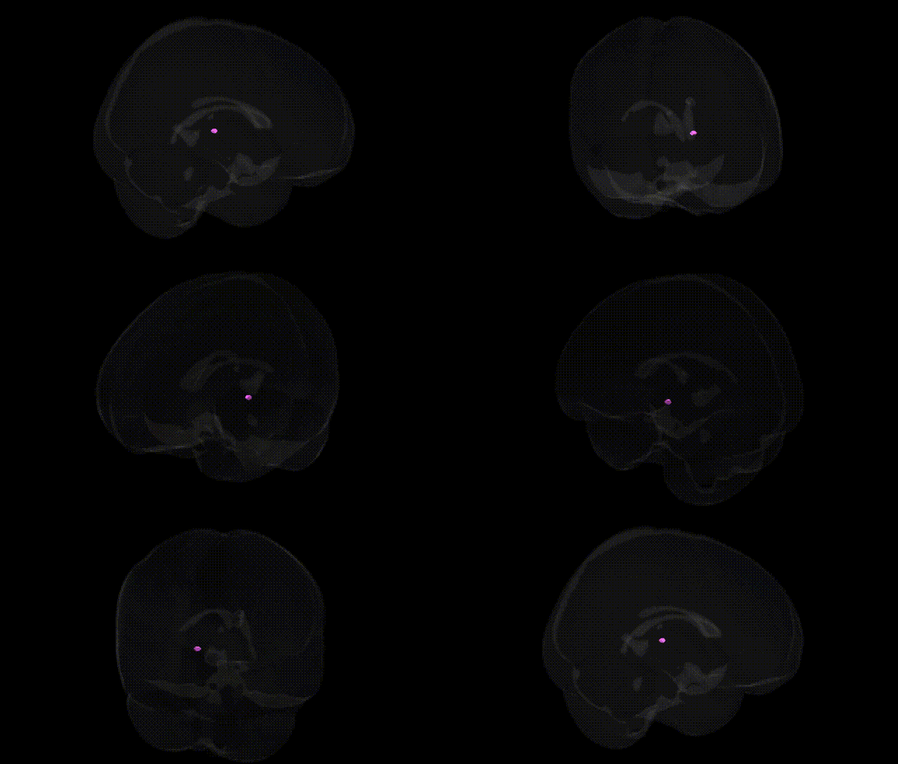
The bilateral Right Brainstem.Midbrain.Gray Matter.Medial Geniculum Body corresponds to the medial geniculate body (MGB), a paired thalamic relay nucleus of the auditory pathway located in the posterior ventral thalamus at the junction of the midbrain and diencephalon. It receives ascending auditory input primarily from the inferior colliculus via the brachium of the inferior colliculus and projects to primary and secondary auditory cortices in the temporal lobe, forming a crucial link for sound processing, including frequency, intensity, and temporal pattern analysis. The MGB is subdivided into ventral, dorsal, and medial divisions, each with distinct cytoarchitecture and connectivity, supporting tonotopic organization and integration of auditory with multisensory and attention-related signals. Lesions in this region can disrupt auditory perception and localization, underscoring its role in conscious hearing and higher-order auditory functions. Medial geniculate nucleus
The medial geniculate body (MGB), the auditory thalamic relay within the midbrain/thalamo–brainstem junction, has been implicated in several genetically influenced traits, although direct GWAS hits explicitly annotated to “medial geniculate” in the Talairach 1 mm atlas are rare and typically embedded within broader thalamic or brainstem measures. Imaging genetics studies and large-scale MRI GWAS (e.g., ENIGMA and UK Biobank–based analyses) have identified associations between polygenic variation and thalamic/brainstem volume or microstructure that encompass the MGB, with notable loci including variants in genes involved in neurodevelopment and axon guidance (such as NRG1, CNTNAP2, and members of the semaphorin and ephrin families) and general neuroanatomical patterning (e.g., loci near HMGA2 for intracranial and subcortical volumes). Functionally, the MGB’s role in auditory processing and sensory gating links it to disorders with auditory or sensory symptoms: candidate-gene and small GWAS-based imaging studies suggest that schizophrenia risk genes (e.g., CACNA1C, GRM3, and COMT pathway variants) may modulate thalamic–midbrain auditory circuitry, contributing to abnormal auditory gating and hallucination-prone phenotypes; autism spectrum disorder and dyslexia risk loci (including genes affecting auditory cortex–thalamus connectivity and language networks) have been associated with altered thalamic and auditory pathway volumes that extend to or include MGB territory; and tinnitus and hyperacusis show emerging, though still inconsistent, genetic signals in ion channel and synaptic genes (e.g., KCNE, GABRA clusters) that likely act partly through midbrain auditory nuclei, including MGB. Additionally, polygenic risk for major depression, anxiety, and attention-deficit/hyperactivity disorder has been correlated with thalamic and brainstem structural variation in GWAS-based imaging studies, implying that common variants in neurodevelopmental and monoaminergic genes influence the structure and function of the auditory thalamus as part of broader cortico–thalamo–brainstem circuits, even though the medial geniculate body is not yet a frequent region-specific label in GWAS summary outputs.
Overview generated by GPT-4o (2026).
Region ID: 499
Hemisphere: bilateral
Atlas: Talairach labels 1mm
Full Quality Version: Download MP4
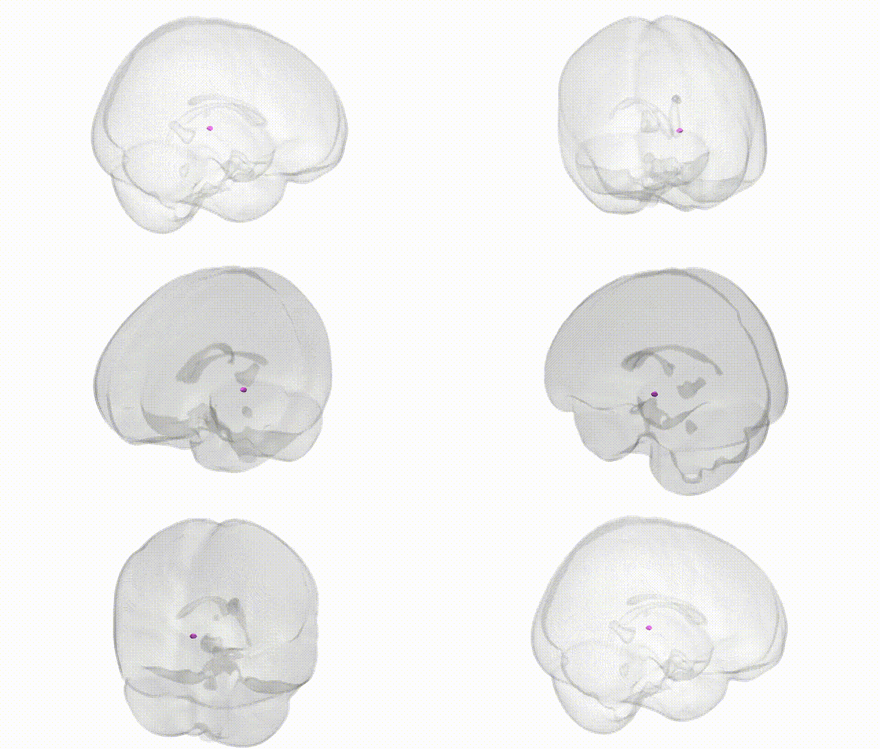
Full Quality Version: Download MP4
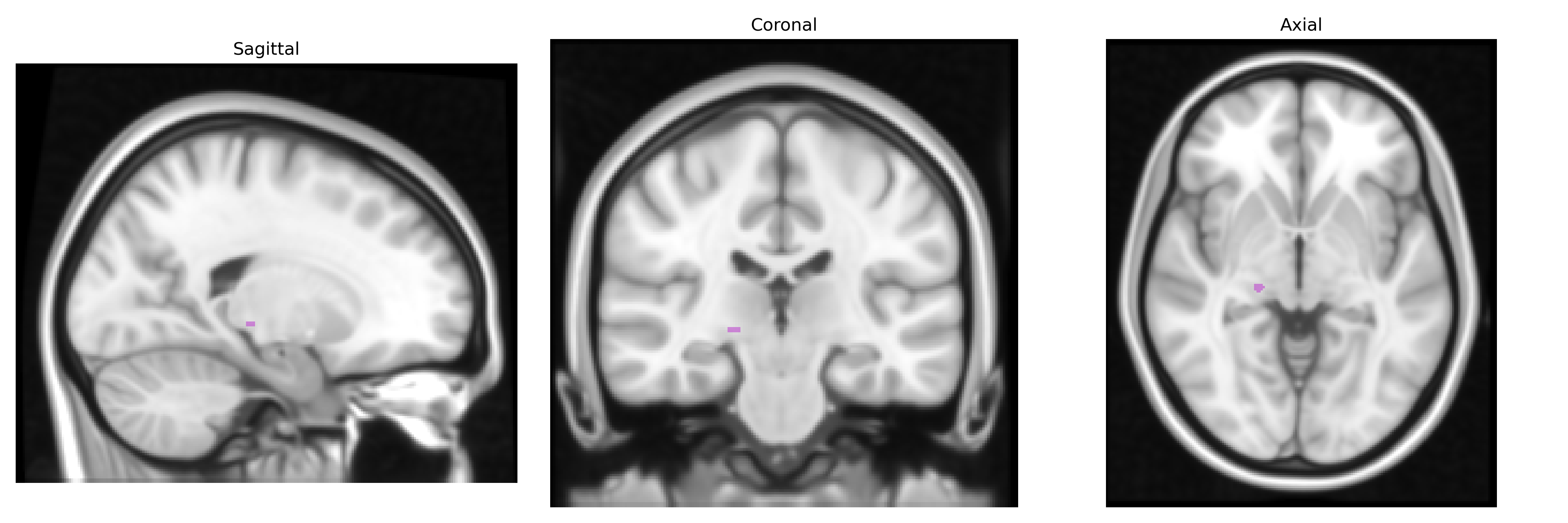
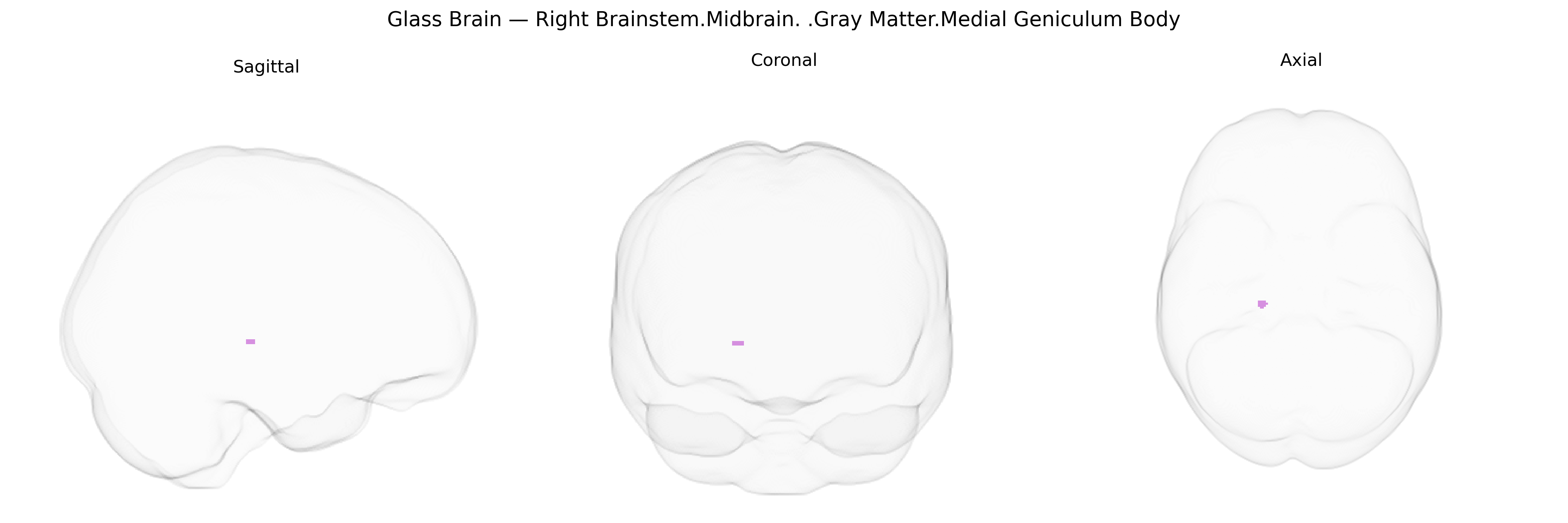
Wali Sidiqyar*, Gaurav Rudravaram*, Elyssa M. McMaster, Trent M. Schwartz, Adam M. Saunders, Kurt G. Schilling, Bennett A. Landman "Introducing SPINS: A Shared Public Visualization Library of Neuroanatomical Structures." Medical Imaging with Deep Learning- short paper
This resource is licensed under CC0 1.0 Universal (Public Domain).