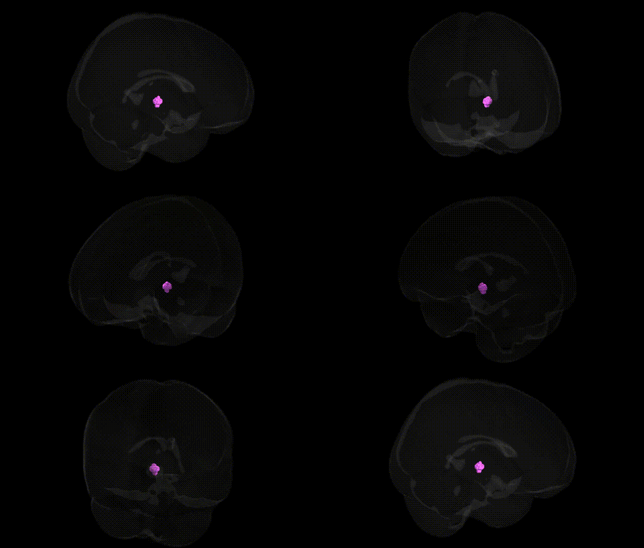
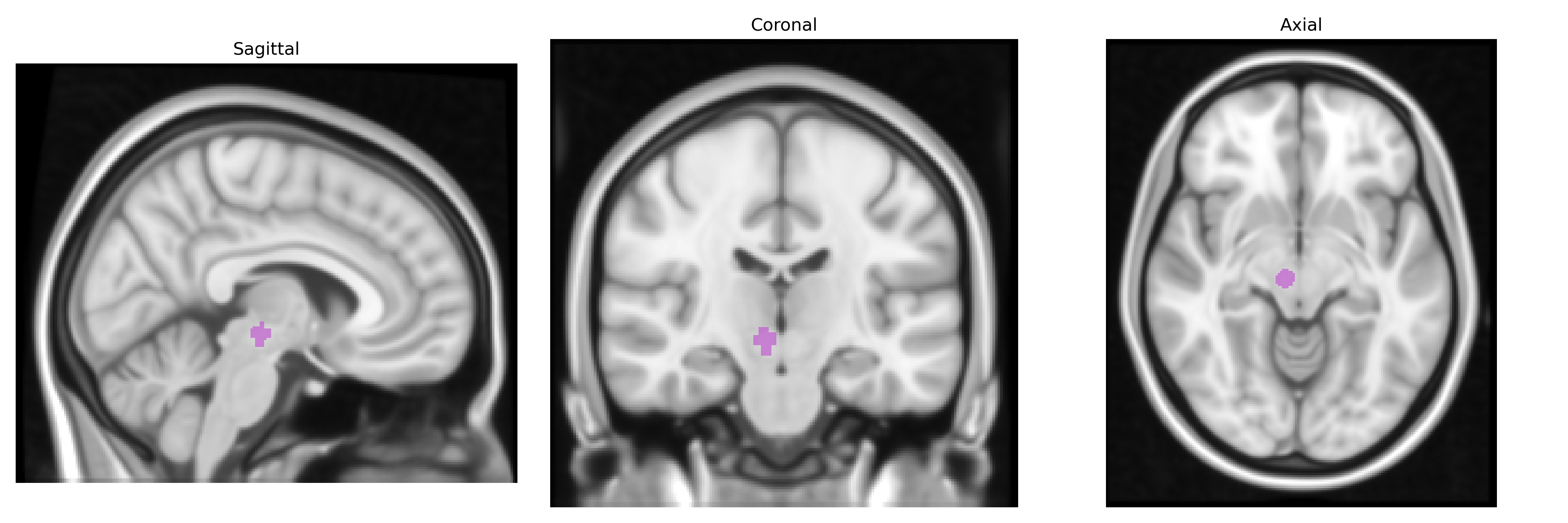
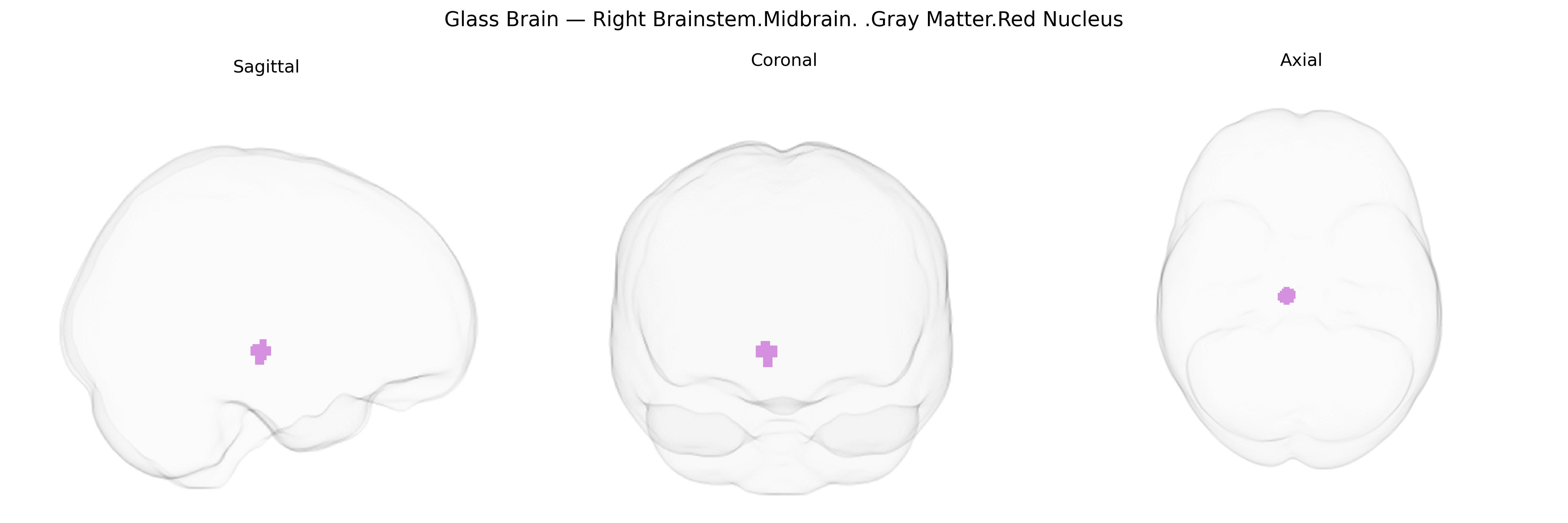
The bilateral Right Brainstem.Midbrain.Gray Matter.Red Nucleus corresponds to a paired, ovoid cluster of magnocellular neurons located in the rostral midbrain tegmentum, medial to the substantia nigra and dorsal to the cerebral peduncles, forming a key relay in the motor circuitry. It receives major afferents from the contralateral cerebellar nuclei and ipsilateral motor cortex, and gives rise to the rubrospinal tract, which influences distal limb flexor musculature and contributes to motor coordination and postural control, particularly in non-human mammals, while in humans its role is more modulatory and integrated with cerebello-thalamo-cortical loops. The red nucleus also participates in motor learning and timing, and its cytoarchitecture and iron-rich neuropil confer the characteristic reddish appearance on gross inspection. Red nucleus
The right midbrain red nucleus, a gray matter structure involved in motor coordination, has limited direct genetic association data, but several GWAS and imaging‑genetics studies implicate genes affecting midbrain and brainstem morphology or function in traits and disorders that plausibly involve this region. Variants in iron‑handling and myelination genes (e.g., HFE, TF, and genes influencing brain iron deposition) have been associated with MRI‑derived iron load in deep brain nuclei, potentially including the red nucleus, which is iron‑rich. Polygenic risk for Parkinson’s disease, involving loci in SNCA, LRRK2, GBA, and MAPT, and dopaminergic pathway genes has been linked to structural and functional alterations in midbrain motor circuits that encompass the red nucleus, as have variants affecting essential tremor and dystonia (e.g., in HTRA2 and TOR1A), although most work focuses on substantia nigra and basal ganglia rather than the red nucleus specifically. Large neuroimaging GWAS (such as those from UK Biobank) have identified common variants in genes related to neurodevelopment, axonal guidance, and synaptic function (e.g., KIAA0319, DLG2, and others) associated with overall brainstem volumes or midbrain morphology, suggesting polygenic influences on this region’s structure, but these findings rarely resolve to the red nucleus at a fine‑grained level like the Talairach 1 mm atlas. Thus, while genetic influences on midbrain and brainstem structure are well supported, specific, replicated GWAS signals tied uniquely to the bilateral right brainstem midbrain gray matter red nucleus region remain sparse, and most genetic links are inferred from broader midbrain or motor‑system imaging and disease studies rather than from region‑exclusive analyses.
Overview generated by GPT-4o (2026).
Region ID: 344
Hemisphere: bilateral
Atlas: Talairach labels 1mm
Full Quality Version: Download MP4
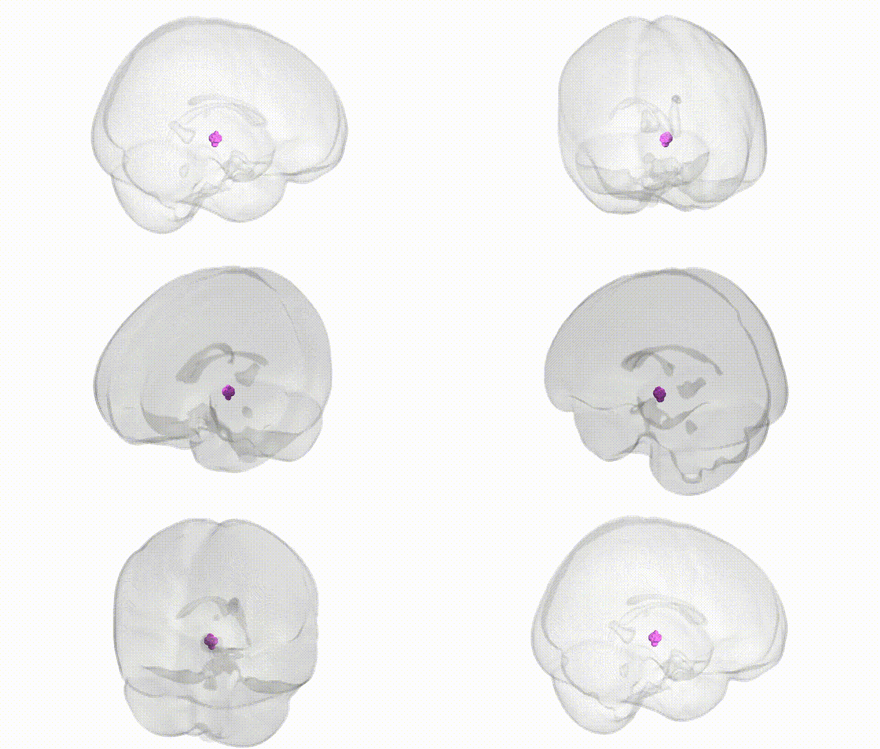
Full Quality Version: Download MP4
Wali Sidiqyar*, Gaurav Rudravaram*, Elyssa M. McMaster, Trent M. Schwartz, Adam M. Saunders, Kurt G. Schilling, Bennett A. Landman "Introducing SPINS: A Shared Public Visualization Library of Neuroanatomical Structures." Medical Imaging with Deep Learning- short paper
This resource is licensed under CC0 1.0 Universal (Public Domain).