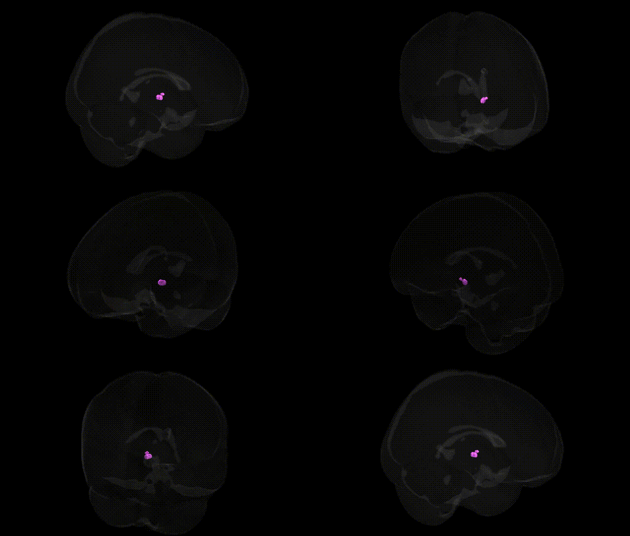
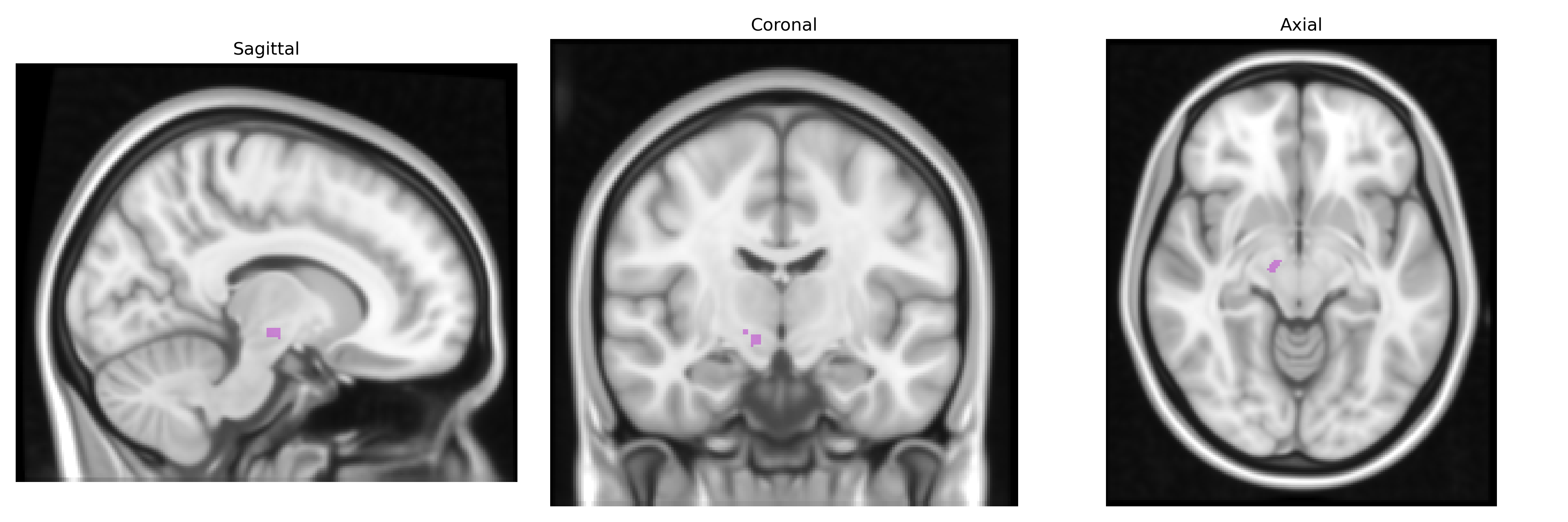
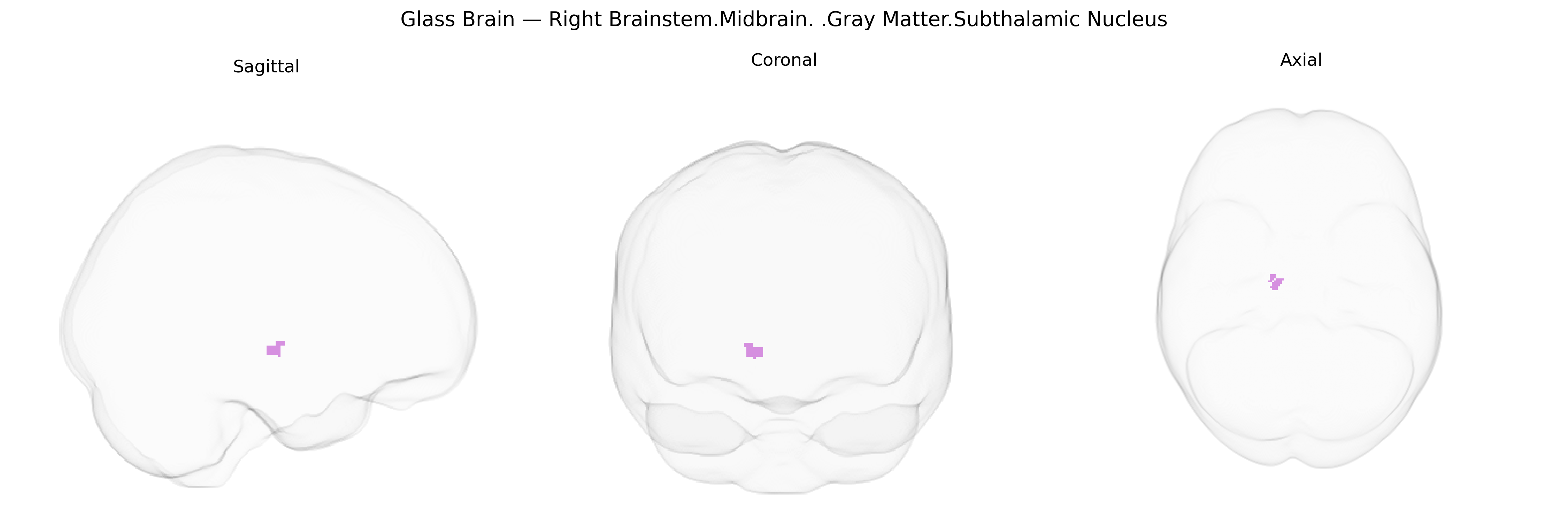
The bilateral Right Brainstem.Midbrain.Gray Matter.Subthalamic Nucleus corresponds to the subthalamic nucleus (STN), a small, lens-shaped gray matter structure located ventral to the thalamus and dorsal to the substantia nigra within the subthalamus of the diencephalon, functionally integrated with the midbrain basal ganglia circuitry. It receives major excitatory glutamatergic input from the cerebral cortex (notably motor areas) via the hyperdirect pathway and inhibitory GABAergic input from the external segment of the globus pallidus, and sends excitatory glutamatergic projections to the internal segment of the globus pallidus and substantia nigra pars reticulata. The STN plays a critical role in modulation of motor output, action selection, and aspects of cognitive and limbic processing; dysfunction or lesion of this nucleus is associated with movement disorders such as hemiballismus and is a key target for deep brain stimulation in Parkinson’s disease. Subthalamic nucleus
The subthalamic nucleus (STN), a key component of the basal ganglia circuitry within the midbrain/brainstem gray matter, has been indirectly implicated in several genetic associations, primarily through disorders and traits characterized by motor and impulse-control dysfunction. Although few GWAS pinpoint the STN with high anatomical specificity, genes strongly linked to Parkinson’s disease (e.g., SNCA, LRRK2, GBA, PARK2) influence neural circuits in which the STN is central, and STN-targeted deep brain stimulation is used to alleviate Parkinsonian motor symptoms that arise from genetically driven dopaminergic and basal ganglia abnormalities. Similarly, variants in genes associated with dystonia (TOR1A), Huntington’s disease (HTT), and rare neurodevelopmental movement disorders alter basal ganglia-thalamo-cortical loops involving the STN, contributing to aberrant motor output and sometimes psychiatric symptoms. In psychiatric genetics, risk variants for obsessive-compulsive disorder and attention-deficit/hyperactivity disorder (e.g., in glutamatergic and dopaminergic signaling genes) have been linked to dysfunction in cortico-striato-thalamo-subthalamic pathways, affecting inhibitory control and compulsivity, though direct STN-focused GWAS evidence remains sparse. Overall, genetic findings point to the STN as a critical relay within circuits affected by disease-associated variants, rather than as a region with many unique, region-specific GWAS hits.
Overview generated by GPT-4o (2026).
Region ID: 454
Hemisphere: bilateral
Atlas: Talairach labels 1mm
Full Quality Version: Download MP4
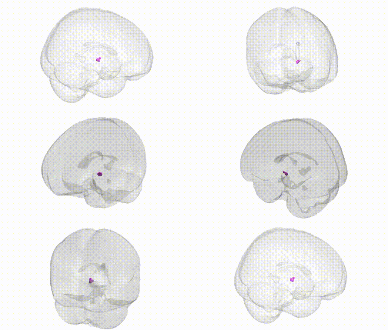
Full Quality Version: Download MP4
Wali Sidiqyar*, Gaurav Rudravaram*, Elyssa M. McMaster, Trent M. Schwartz, Adam M. Saunders, Kurt G. Schilling, Bennett A. Landman "Introducing SPINS: A Shared Public Visualization Library of Neuroanatomical Structures." Medical Imaging with Deep Learning- short paper
This resource is licensed under CC0 1.0 Universal (Public Domain).