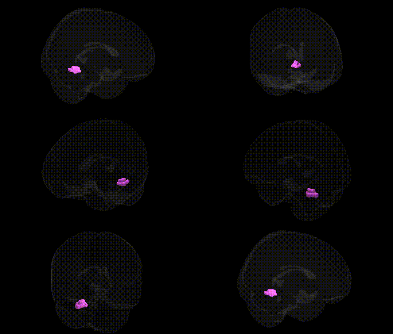
The bilateral Right Cerebellum, Anterior Lobe, Gray Matter, Dentate region corresponds to the dentate nucleus located within the anterior lobe of the right cerebellar hemisphere, representing a major deep cerebellar nucleus composed of densely packed gray matter. This structure plays a central role in the coordination and fine-tuning of voluntary movements, motor planning, and the integration of sensory and motor information, receiving input from Purkinje cells of the cerebellar cortex and projecting via the superior cerebellar peduncle to motor and premotor areas of the thalamus and cerebral cortex. Functionally, it contributes to precision, timing, and learning of movements and may also participate in certain cognitive and language-related processes via its extensive cerebello-thalamo-cortical connections. There is no direct link for this composite Talairach label; a related structure is the Dentate nucleus.
The bilateral right cerebellar anterior lobe gray matter of the dentate nucleus (as defined in Talairach 1 mm labels) participates in motor coordination, timing, and cognitive–affective processing, and genetic associations involving this region arise largely from imaging genetics and cerebellar volume GWAS rather than region-specific candidate gene studies. Large-scale MRI–GWAS datasets (e.g., UK Biobank–based analyses) have identified common variants in genes implicated in neurodevelopment, synaptic function, and axon guidance (such as variants in MAPT, GRIN2B, and neurodevelopmental loci near FOXP1 and LINGO1) that show associations with cerebellar gray matter volume and morphology, including dentate and anterior lobe territories. Polygenic architectures for traits like general cognitive ability, educational attainment, and motor performance display genetic correlations with cerebellar structure, and several schizophrenia, bipolar disorder, and major depression risk loci exhibit pleiotropic effects on cerebellar volumes or functional connectivity involving dentate regions in imaging–genetics analyses. Additionally, rare variants and copy-number changes affecting genes involved in cerebellar development (e.g., in autosomal recessive cerebellar ataxias and some autism spectrum or intellectual disability genes such as RELN and ASTN2) can lead to structural and volumetric alterations encompassing the anterior lobe and dentate nucleus. Parkinson’s disease and essential tremor GWAS have also implicated loci (for example near LINGO1 and STK32B) that, through downstream imaging studies, have been associated with altered cerebellar–dentate structure or function, supporting a genetically mediated link between this region, movement disorders, and broader neuropsychiatric and cognitive traits, although no single locus is uniquely specific to the “Right Cerebellum.Anterior Lobe.Gray Matter.Dentate” label as currently defined.
Overview generated by GPT-4o (2026).
Region ID: 118
Hemisphere: bilateral
Atlas: Talairach labels 1mm
Full Quality Version: Download MP4
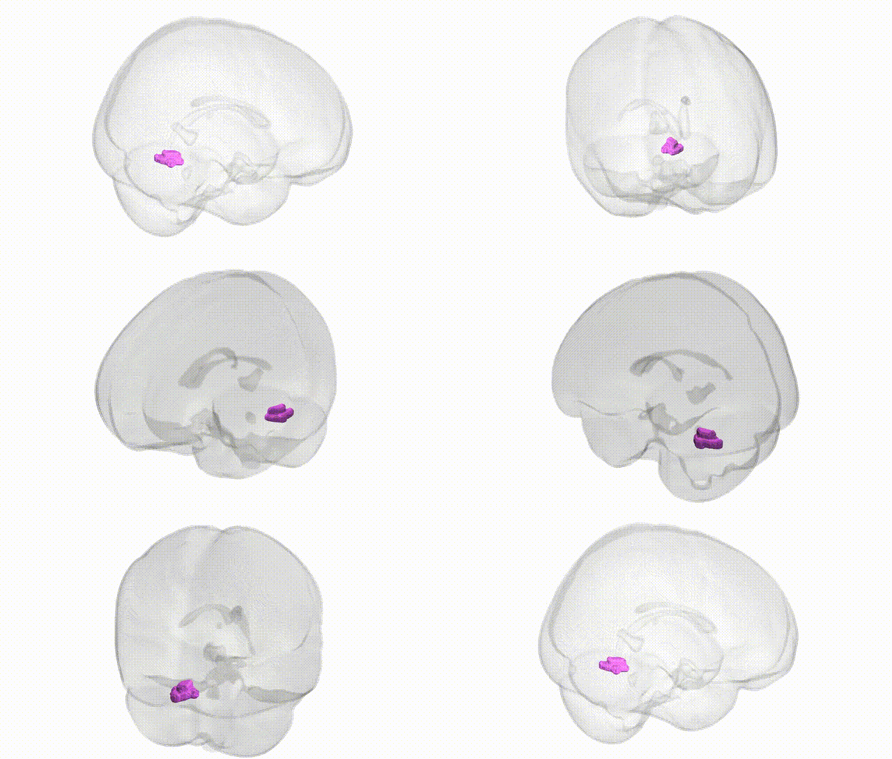
Full Quality Version: Download MP4
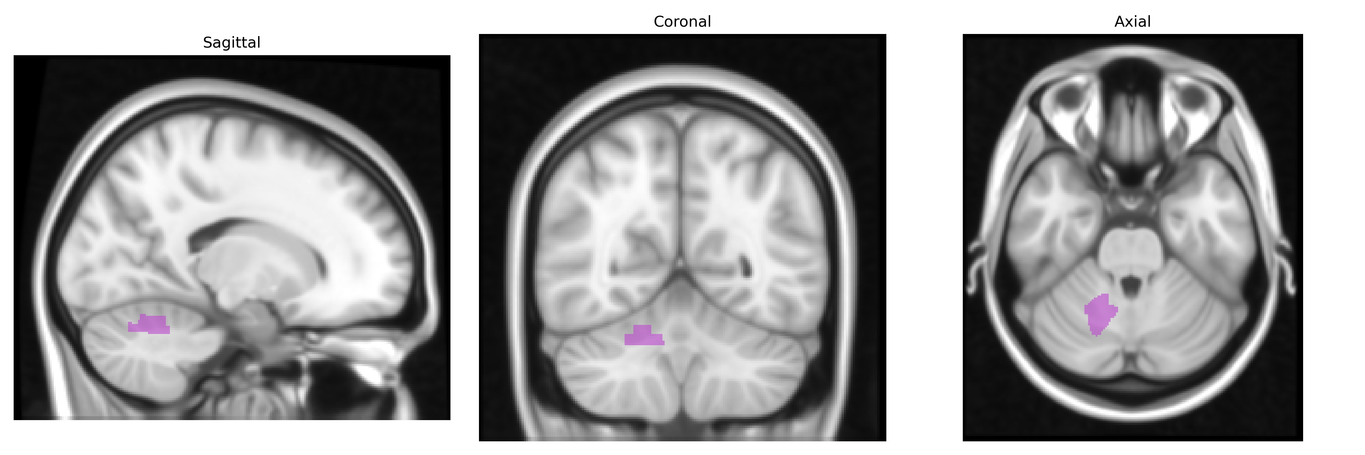
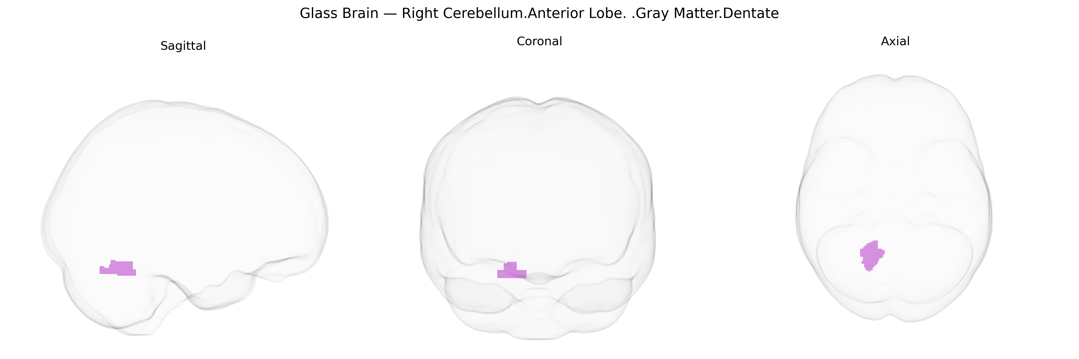
Wali Sidiqyar*, Gaurav Rudravaram*, Elyssa M. McMaster, Trent M. Schwartz, Adam M. Saunders, Kurt G. Schilling, Bennett A. Landman "Introducing SPINS: A Shared Public Visualization Library of Neuroanatomical Structures." Medical Imaging with Deep Learning- short paper
This resource is licensed under CC0 1.0 Universal (Public Domain).