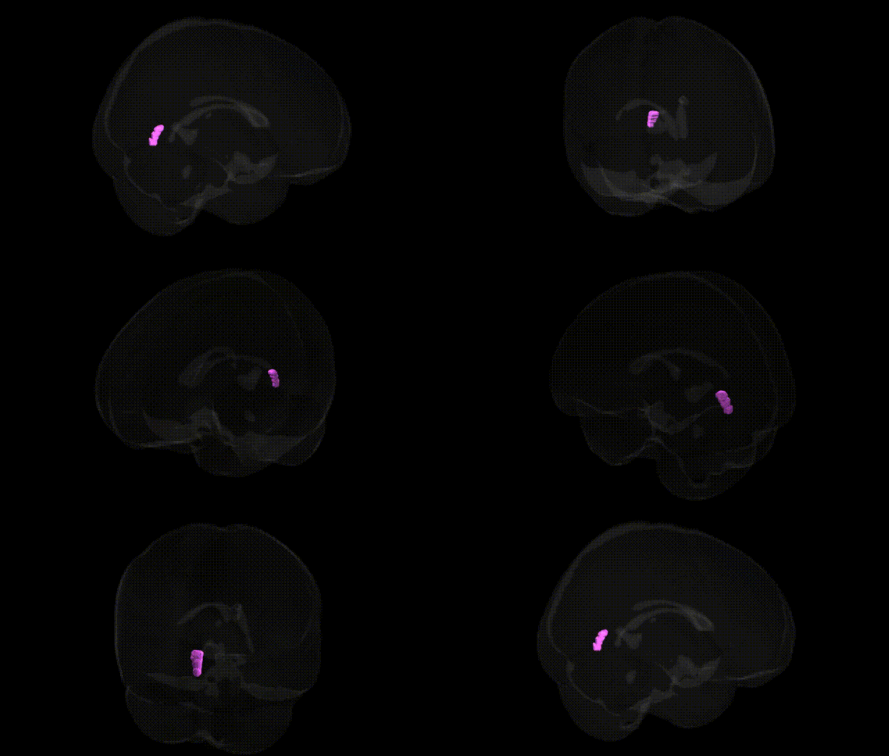
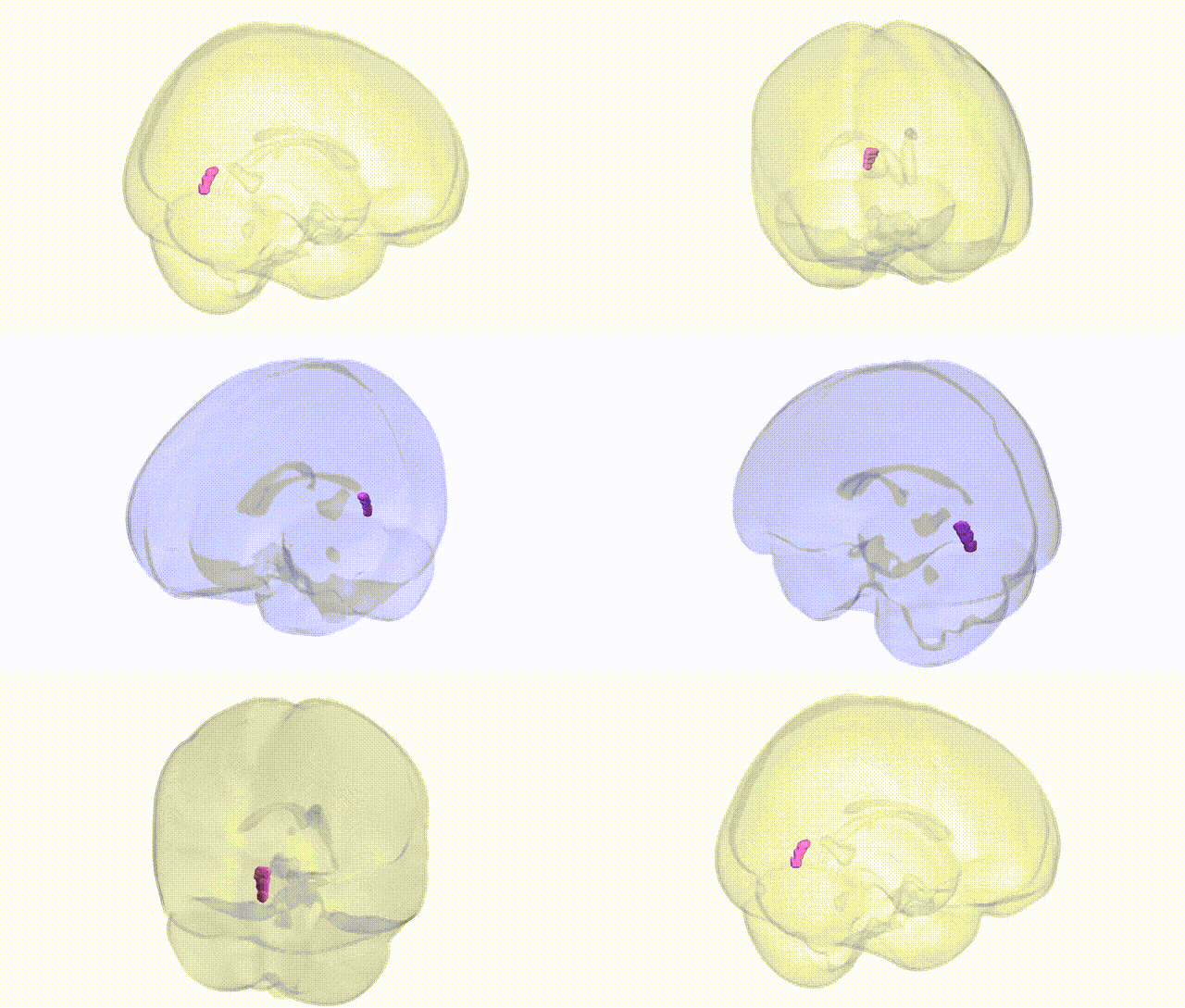
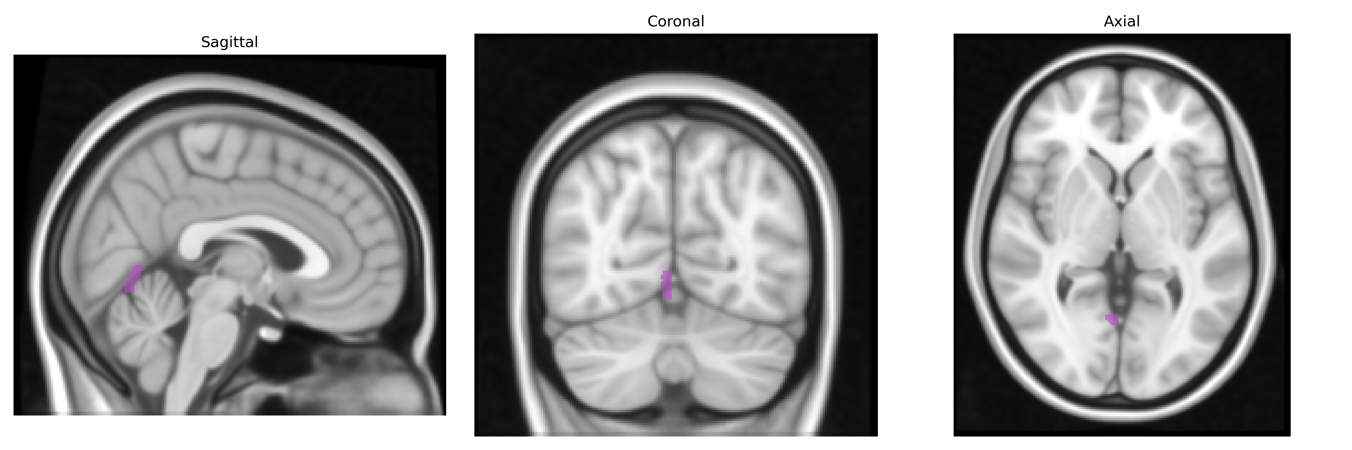
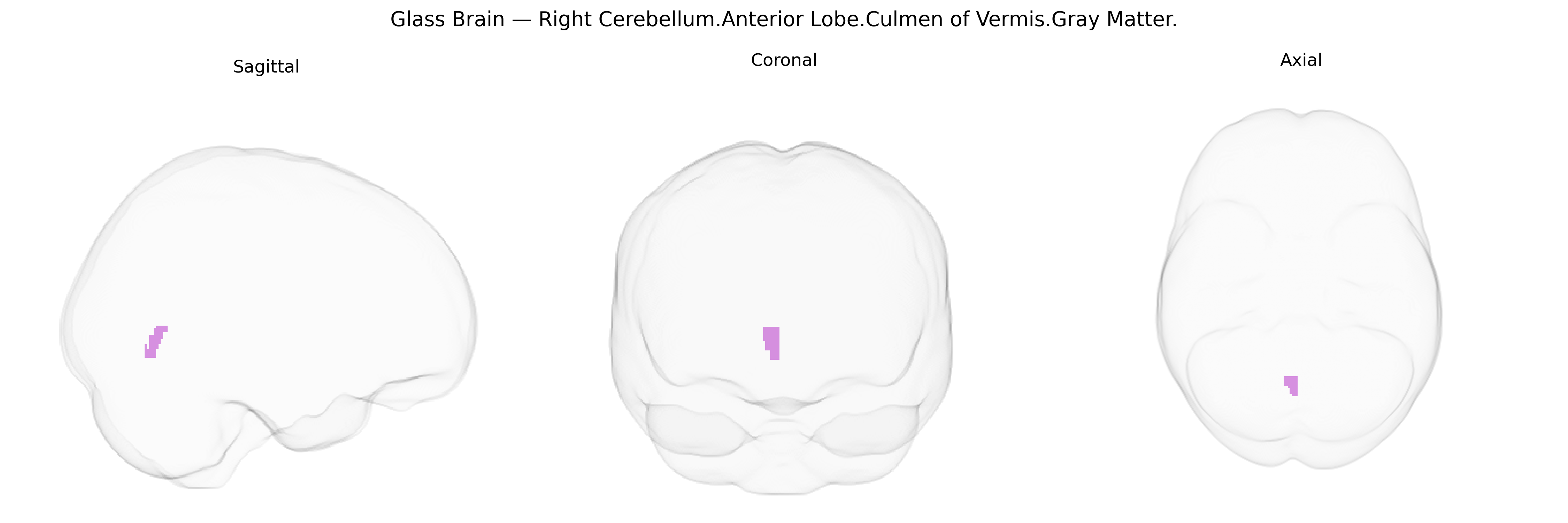
The bilateral Right Cerebellum.Anterior Lobe.Culmen of Vermis.Gray Matter corresponds to gray matter within the culmen, a lobule of the anterior lobe of the cerebellar vermis located on the dorsal aspect of the cerebellum. This region participates in sensorimotor integration, particularly in the coordination of posture, gait, and fine-tuned limb movements, receiving somatosensory input and contributing to the modulation of motor output via cerebellar cortical circuits projecting to deep cerebellar nuclei. Functionally, it is involved in error correction during movement, helping to maintain balance and adapt motor patterns based on feedback, and may also contribute to aspects of motor learning. Histologically, this gray matter displays the characteristic cerebellar cortical layering (molecular, Purkinje cell, and granular layers), supporting high-density synaptic processing of afferent inputs from spinal, vestibular, and cortical sources. Related article: Cerebellar vermis.
The Right Cerebellum Anterior Lobe (Culmen of Vermis, gray matter) in the Talairach 1 mm atlas corresponds largely to lobules IV–V of the anterior cerebellum, a region implicated in motor coordination, balance, and aspects of cognitive and affective processing, and its genetic associations are typically inferred from broader cerebellar or lobular GWAS rather than this precise parcel. Large-scale imaging–genetics studies (e.g., UK Biobank–based GWAS of regional brain volumes and cortical/cerebellar morphology) have identified multiple loci influencing anterior cerebellar volume and vermis structure, including variants near genes involved in neurodevelopment and axon guidance such as MAPT, PLA2G4E, and several chromatin-remodeling and synaptic genes, though mapping to the specific Culmen of Vermis label is indirect. GWAS of neurodevelopmental and psychiatric disorders that show structural or functional alterations of the anterior cerebellar vermis—such as autism spectrum disorder, schizophrenia, attention-deficit/hyperactivity disorder, and bipolar disorder—implicate polygenic risk across synaptic, calcium-signaling, and neurodevelopmental pathways (e.g., genes like CACNA1C, GRIN2A, and multiple glutamatergic and GABAergic genes), with imaging–genetic mediation analyses suggesting that cerebellar vermis morphology can partially bridge genetic risk and clinical phenotype in some cohorts. In movement and coordination disorders, rare and common variants in genes linked to spinocerebellar ataxias (e.g., ATXN1, ATXN2, ATXN3, CACNA1A) and other cerebellar atrophy syndromes often show early involvement of anterior cerebellar and vermal regions, including the culmen, although these data usually derive from clinical and structural imaging rather than voxelwise atlas-based mapping. Pain, balance, and motor-control traits in GWAS have also been associated with cerebellar volume measures, but the specific Culmen of Vermis region appears as part of broader anterior cerebellar clusters. Overall, current genetic evidence connects polygenic neurodevelopmental and synaptic pathways to anterior cerebellar and vermal structure and function, with this atlas-defined region participating in those associations rather than being isolated as a uniquely and independently identified genetic target.
Overview generated by GPT-4o (2026).
Region ID: 322
Hemisphere: bilateral
Atlas: Talairach labels 1mm
Full Quality Version: Download MP4
Full Quality Version: Download MP4
Wali Sidiqyar*, Gaurav Rudravaram*, Elyssa M. McMaster, Trent M. Schwartz, Adam M. Saunders, Kurt G. Schilling, Bennett A. Landman "Introducing SPINS: A Shared Public Visualization Library of Neuroanatomical Structures." Medical Imaging with Deep Learning- short paper
This resource is licensed under CC0 1.0 Universal (Public Domain).