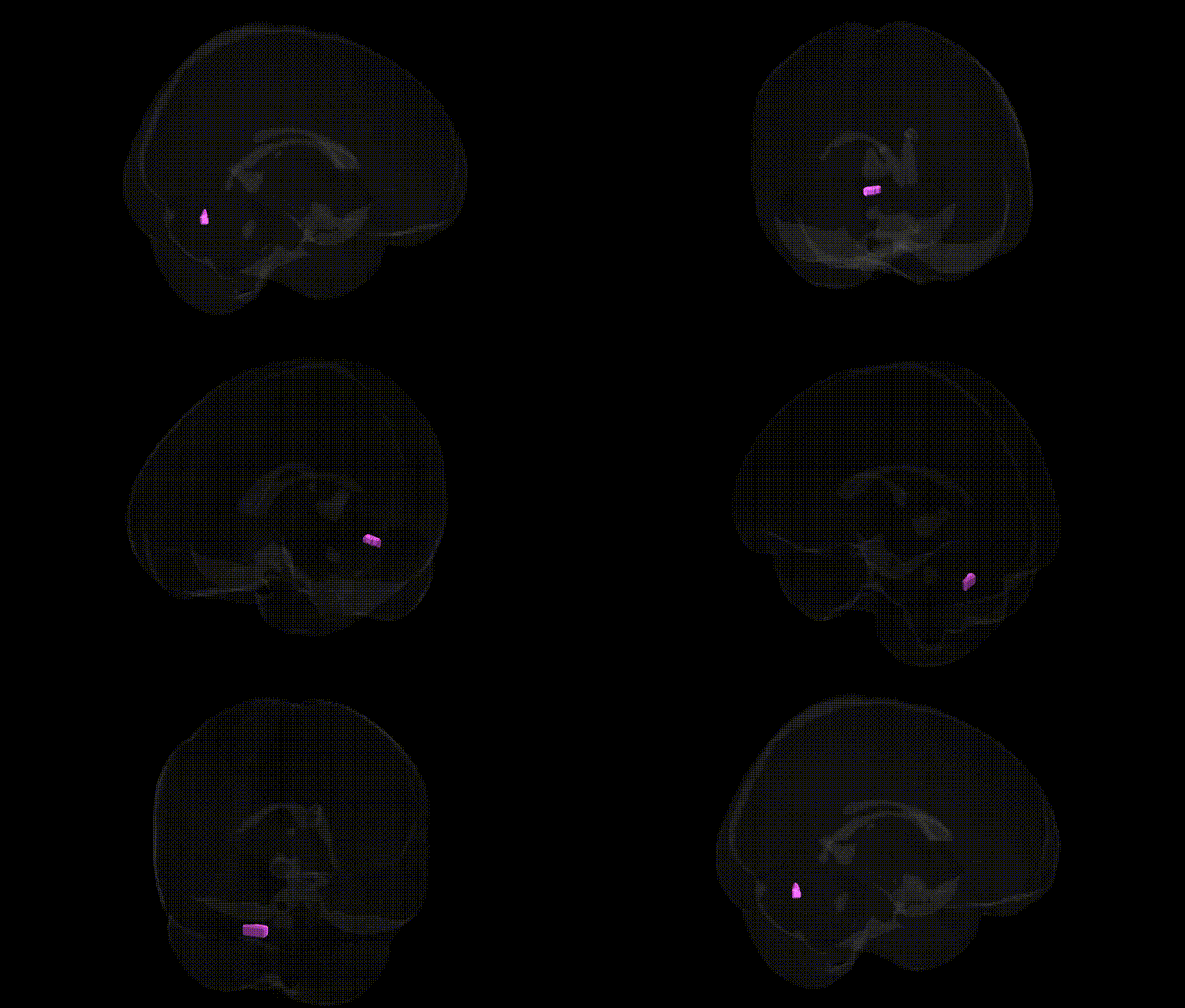
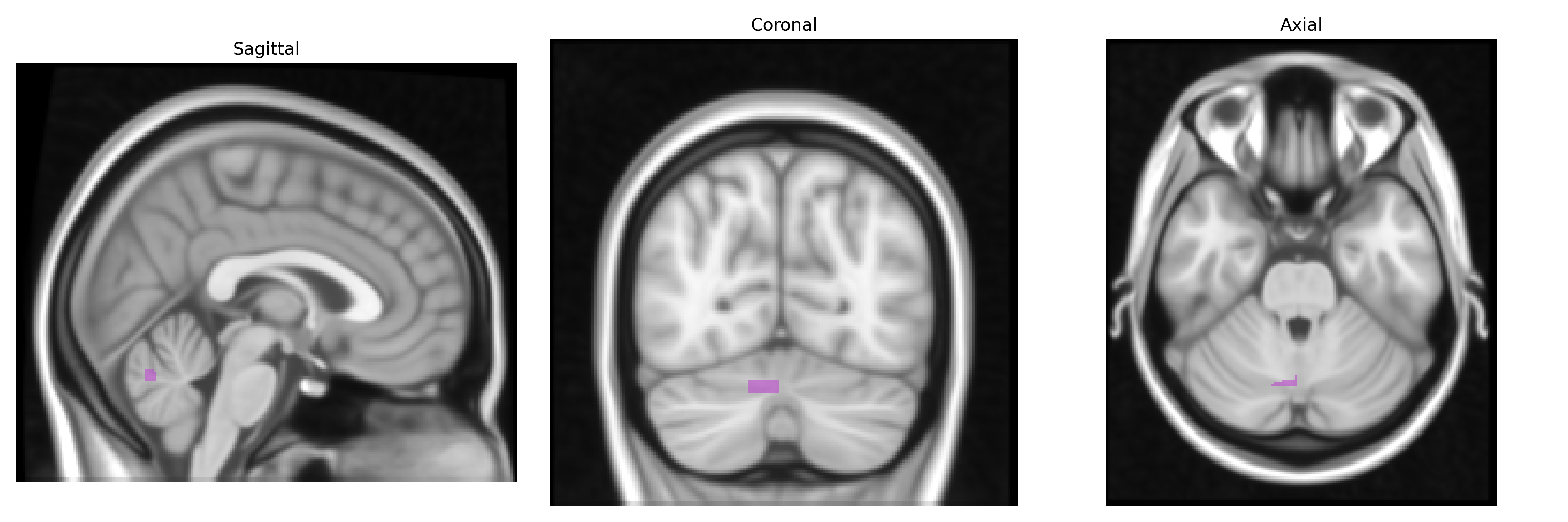
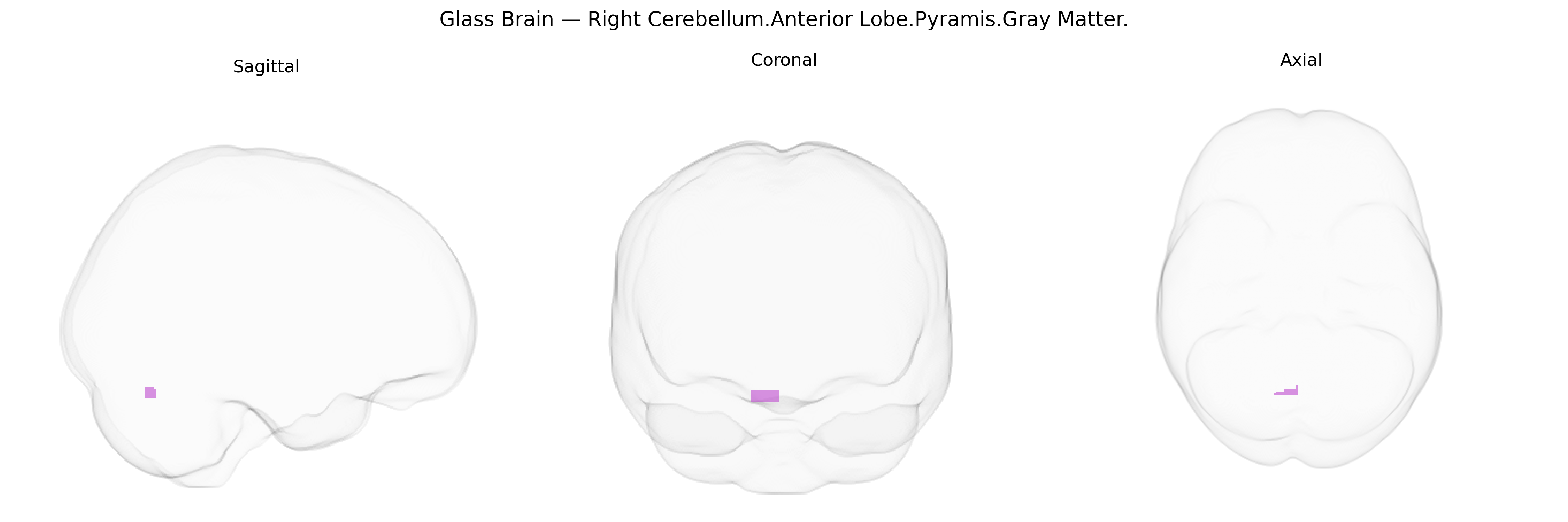
The bilateral Right Cerebellum.Anterior Lobe.Pyramis.Gray Matter refers to gray matter within the pyramis of the anterior lobe of the right cerebellar hemisphere, a vermal structure that contributes to the regulation of posture, muscle tone, and coordination of axial and proximal limb movements. This region is part of the spinocerebellum, receiving somatosensory input from the spinal cord and projecting primarily to fastigial and interposed cerebellar nuclei, which in turn influence vestibulospinal, reticulospinal, and other descending motor pathways. Functionally, it participates in error correction during ongoing movements and in the fine-tuning of balance and stance, integrating sensory feedback to adjust motor output. There is no direct link for this specific subregion; a related article is: Cerebellum.
The bilateral right cerebellar anterior lobe (pyramis, gray matter) as defined in the Talairach 1 mm atlas has not featured prominently as a primary locus in large-scale GWAS, but genetic studies implicating cerebellar volume, structure, and connectivity suggest indirect associations with this region. Polygenic influences on total cerebellar and lobular volumes have been linked to variants in genes involved in neurodevelopment (e.g., KIRREL3, WNT and FGF pathway genes, and axon guidance genes) and synaptic organization, with several GWAS of brain MRI phenotypes showing cerebellar lobules among the heritable structural traits, though typically at the level of broad anterior/posterior cerebellar regions rather than the specific pyramis. Cerebellar gray matter alterations involving the anterior lobe, including the vermal and paravermal pyramis, have been reported in neurodevelopmental and psychiatric disorders such as autism spectrum disorder, schizophrenia, bipolar disorder, and major depression, where associated risk variants commonly involve synaptic plasticity (e.g., CACNA1C, GRIN2A/B), neurodevelopmental regulators (e.g., CNTNAP2, DISC1), and immune or microglial function genes, suggesting that polygenic risk for these conditions may contribute to structural and functional variability in this region. In movement and coordination disorders, including spinocerebellar ataxias (e.g., expansions in ATXN1, ATXN2, ATXN3, and CACNA1A mutations), volumetric and degenerative changes are widespread but often include the anterior cerebellar lobules and the pyramis, linking specific monogenic variants to pathology in this territory. Additionally, GWAS of motor, balance, and cognitive traits, as well as imaging-genetics studies from resources like UK Biobank, have reported associations between polygenic scores for cognitive performance, educational attainment, and psychiatric risk and variation in cerebellar gray matter and connectivity, consistent with a role for genetically influenced cerebellar circuits—including the anterior lobe and pyramis—in motor control, timing, and higher-order cognitive and affective processes, even though precise gene–region mappings at the resolution of “Right Cerebellum.Anterior Lobe.Pyramis.GRAY” remain limited.
Overview generated by GPT-4o (2026).
Region ID: 117
Hemisphere: bilateral
Atlas: Talairach labels 1mm
Full Quality Version: Download MP4
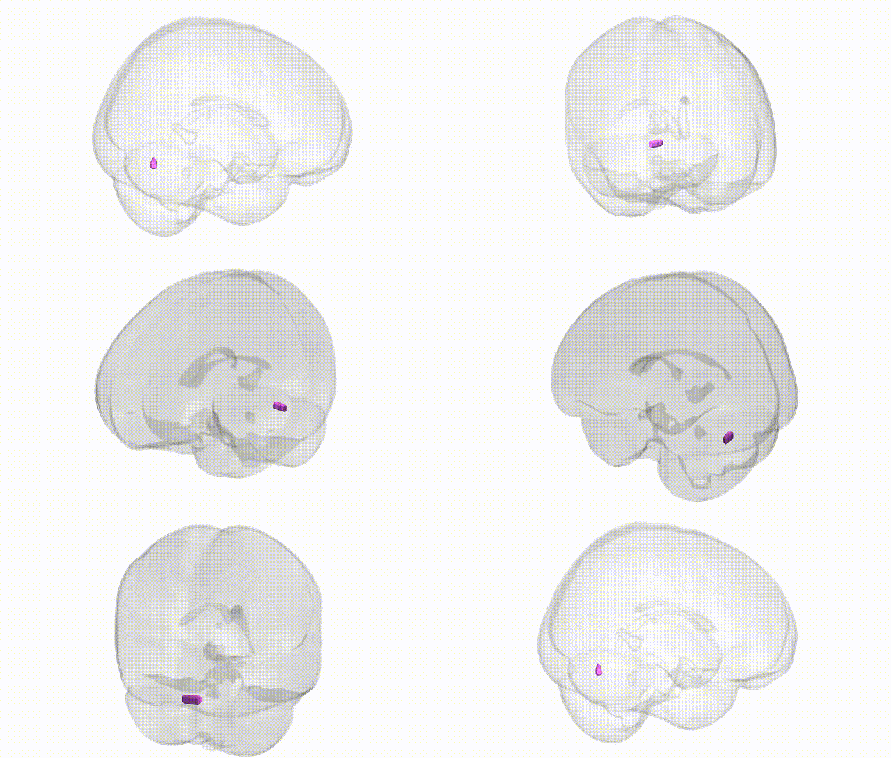
Full Quality Version: Download MP4
Wali Sidiqyar*, Gaurav Rudravaram*, Elyssa M. McMaster, Trent M. Schwartz, Adam M. Saunders, Kurt G. Schilling, Bennett A. Landman "Introducing SPINS: A Shared Public Visualization Library of Neuroanatomical Structures." Medical Imaging with Deep Learning- short paper
This resource is licensed under CC0 1.0 Universal (Public Domain).