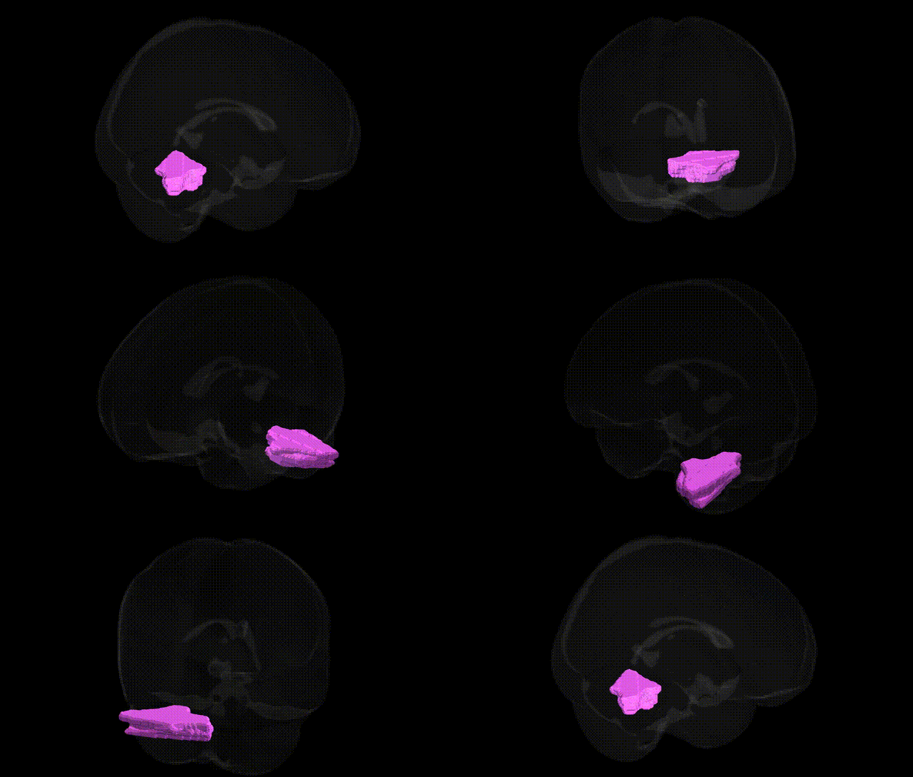
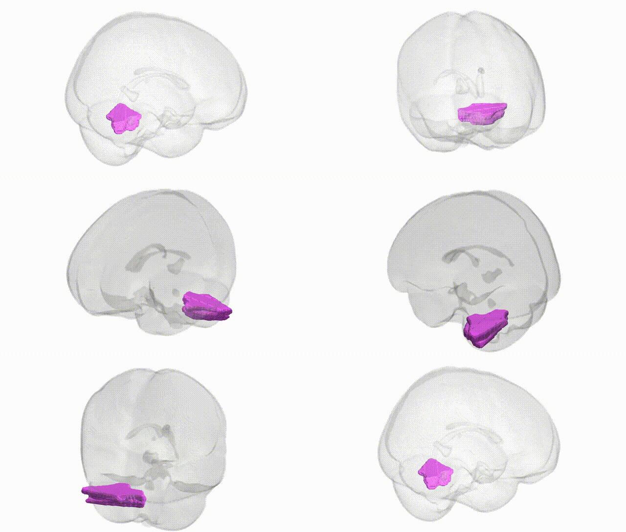
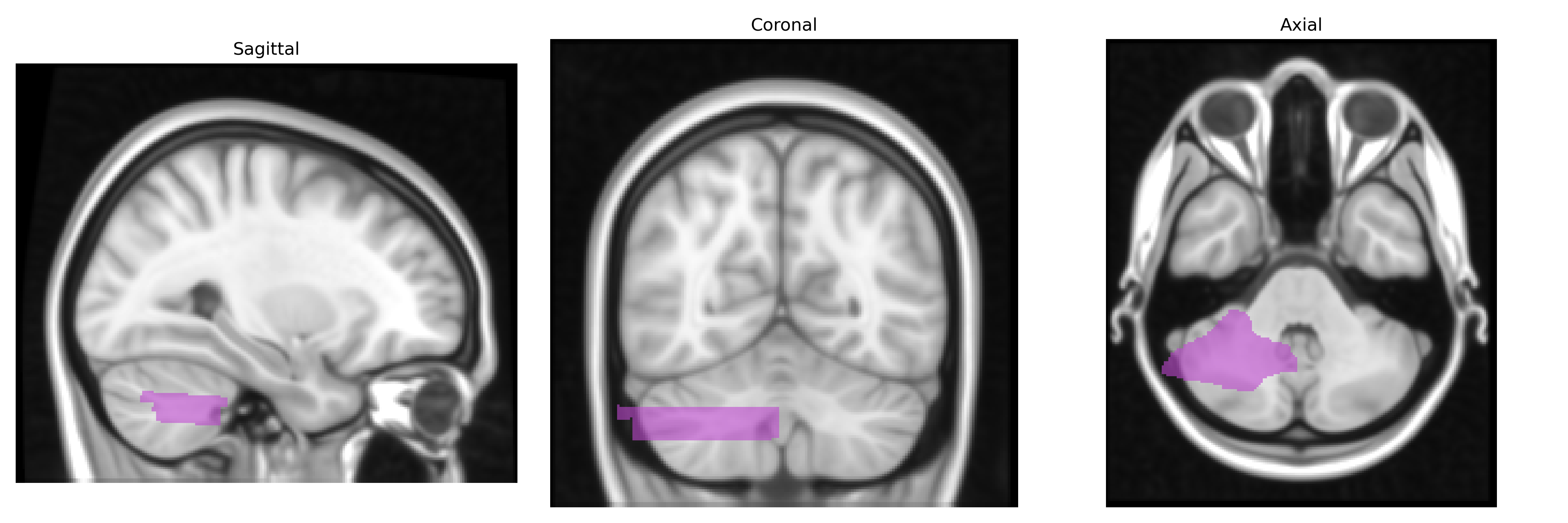
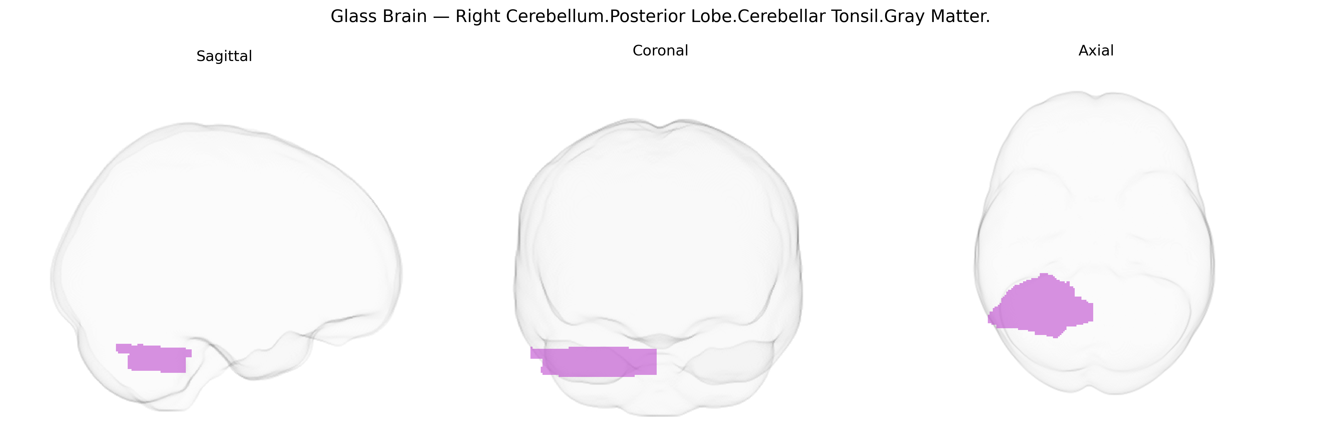
The bilateral Right Cerebellum.Posterior Lobe.Cerebellar Tonsil.Gray Matter corresponds to the gray-matter portions of the cerebellar tonsils located in the inferior aspect of the posterior lobe of the cerebellum, adjacent to the medulla and near the foramen magnum. These ovoid masses of cortex and underlying nuclei participate in fine motor coordination, balance, and the modulation of muscle tone, integrating sensory and motor signals from spinal, vestibular, and cortical inputs. Anatomically, the cerebellar tonsils are significant for their propensity to herniate caudally through the foramen magnum in posterior fossa crowding or Chiari malformations, potentially compressing the medulla and disrupting cardiorespiratory and motor functions. There is no direct link for this exact composite label; see the related structure: Cerebellar tonsil.
The bilateral right cerebellar tonsil of the posterior lobe (gray matter), as defined in the Talairach 1 mm atlas, has been implicated in genetic studies largely through broader cerebellar and posterior lobe measures rather than highly specific tonsillar loci, with several GWAS identifying heritable influences on cerebellar volume and morphology that encompass this region. Large-scale imaging-genetics consortia (e.g., ENIGMA, UK Biobank) report polygenic contributions to cerebellar structure, with common variants in genes involved in neurodevelopment, axon guidance, synaptic function, and neuronal migration (such as those inRELN,ROBO, and neurodevelopmental pathways) contributing to interindividual differences in posterior cerebellar gray matter volumes that include the tonsils. Rare-variant and linkage studies in congenital cerebellar malformations (e.g., Chiari I malformation, Dandy–Walker spectrum) occasionally highlight structural anomalies involving the cerebellar tonsils, with associated genes in hindbrain patterning, cranial mesoderm, and connective tissue (including collagen and extracellular matrix genes) playing a role, although findings remain heterogeneous and often not region-specific. Neuropsychiatric and neurodevelopmental GWAS (in disorders such as schizophrenia, autism spectrum disorder, ADHD, and mood disorders) consistently show polygenic overlap with global and regional cerebellar volumes, including posterior lobe territories, implicating shared genetic architectures rather than single loci uniquely tied to the tonsil. Overall, current evidence supports a polygenic, system-level genetic influence on cerebellar posterior lobe and tonsillar gray matter, with specific gene–region associations still limited and typically inferred from broader cerebellar volumetric and malformation-related genetic studies rather than isolated GWAS signals focused exclusively on the right cerebellar tonsil.
Overview generated by GPT-4o (2026).
Region ID: 4
Hemisphere: bilateral
Atlas: Talairach labels 1mm
Full Quality Version: Download MP4
Full Quality Version: Download MP4
Wali Sidiqyar*, Gaurav Rudravaram*, Elyssa M. McMaster, Trent M. Schwartz, Adam M. Saunders, Kurt G. Schilling, Bennett A. Landman "Introducing SPINS: A Shared Public Visualization Library of Neuroanatomical Structures." Medical Imaging with Deep Learning- short paper
This resource is licensed under CC0 1.0 Universal (Public Domain).