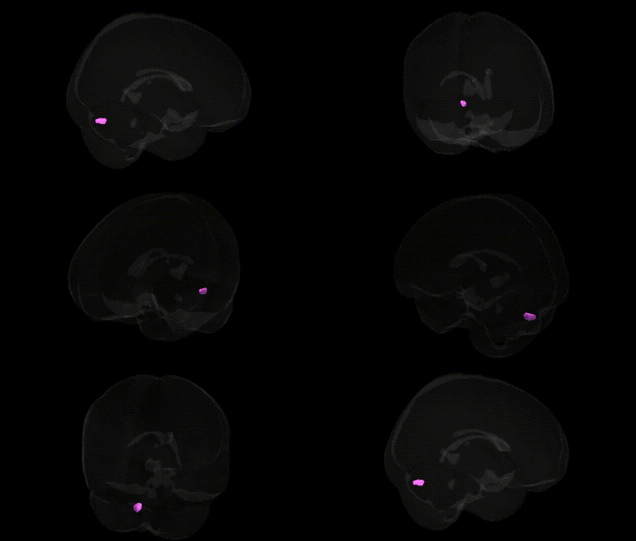
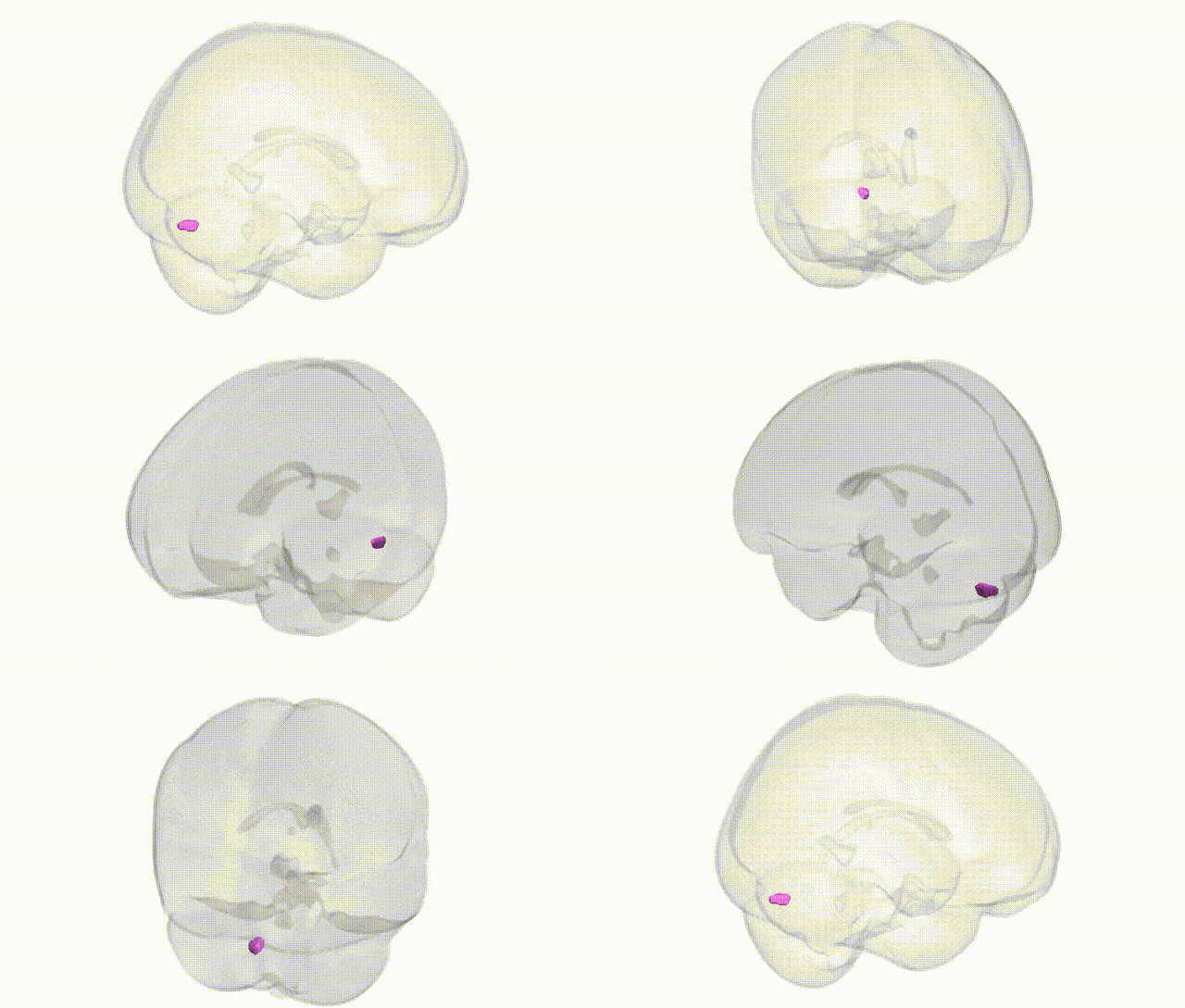
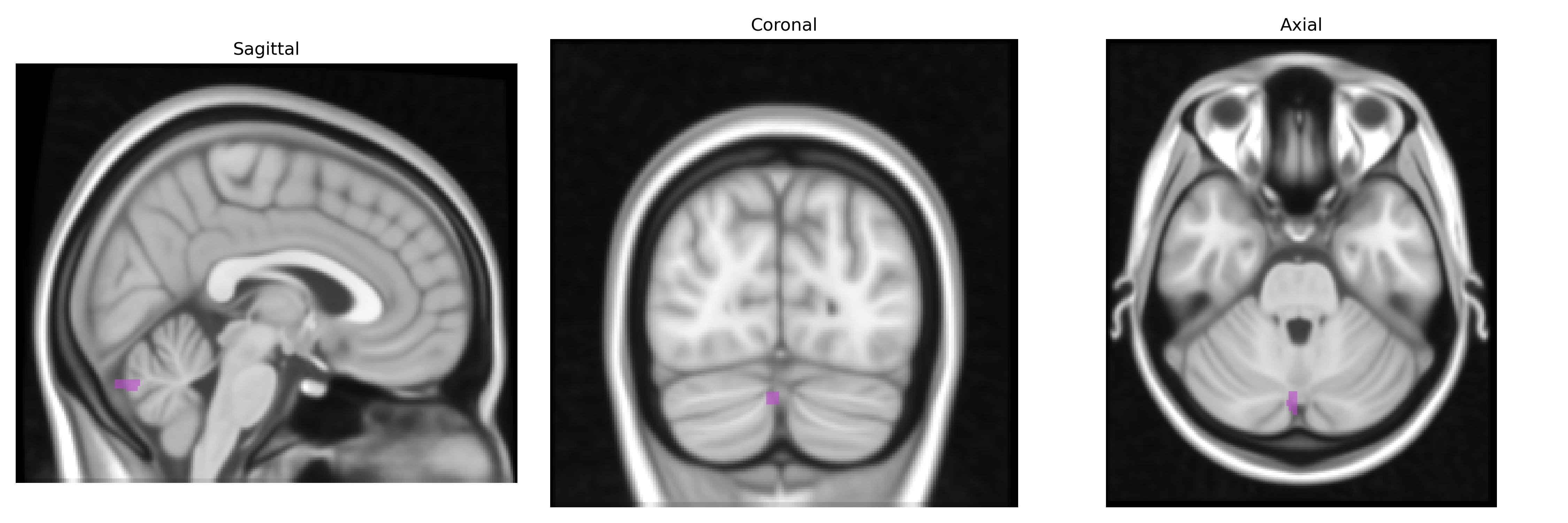
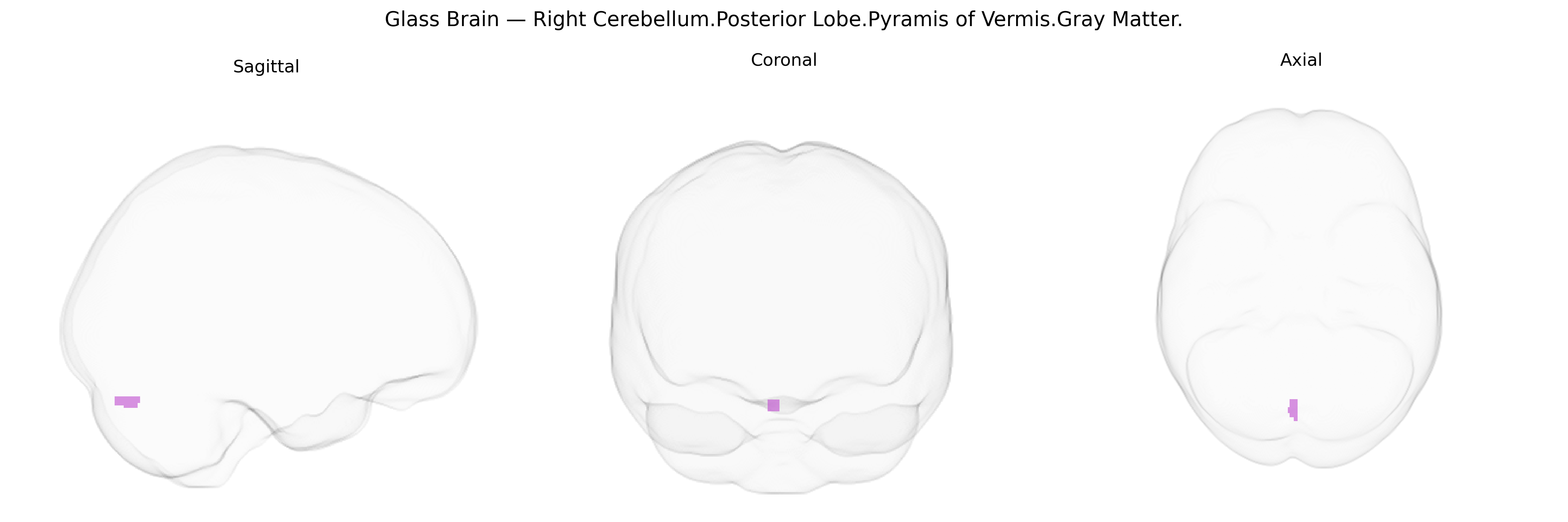
The bilateral Right Cerebellum Posterior Lobe Pyramis of Vermis Gray Matter corresponds to a midline structure of the cerebellar vermis located within the posterior lobe, composed predominantly of cortical gray matter that overlays the deep cerebellar white matter. The pyramis participates in the regulation of axial and proximal limb musculature and contributes to postural control, balance, and coordination of movements through its connections with spinocerebellar pathways and vestibular and reticular nuclei. Functionally, it is considered part of the spinocerebellum, integrating somatosensory feedback to fine-tune ongoing motor activity and maintain equilibrium. There is no direct link for the pyramis of the vermis; a related structure is the cerebellar vermis: Cerebellar vermis
The bilateral right cerebellar posterior lobe, specifically the pyramis of the vermis gray matter, has been implicated in genetic studies largely through imaging genetics and cerebellar volume GWAS rather than region-specific loci unique to the Talairach 1 mm atlas label. Large-scale GWAS of cerebellar structure and total cerebellar volume (e.g., ENIGMA and UK Biobank–based studies) have identified multiple associated loci in genes involved in neurodevelopment, synaptic function, and axonal guidance (such as FOXP1/FOXP2 regions, GRID2, and variants near MAPT and AUTS2), with some associations overlapping with global or lobule-specific measures that include posterior vermis territories. Genetic correlations and pleiotropic analyses show that cerebellar volume and regional morphology exhibit shared genetic architecture with psychiatric and neurodevelopmental conditions, including schizophrenia, bipolar disorder, major depressive disorder, autism spectrum disorder, and ADHD, as well as cognitive traits and general intelligence, suggesting that variants influencing cerebellar posterior lobe and vermal structure may contribute to vulnerability for these disorders. Additional work in spinocerebellar ataxias (e.g., expansions in ATXN1–3, CACNA1A) and related hereditary degenerative disorders consistently demonstrates pronounced atrophy in midline vermis structures (including the pyramis), linking pathogenic repeat expansions and ion channel genes to degeneration in this region, although these are typically rare, high-penetrance mutations rather than common GWAS variants. Overall, genetic findings converge on the cerebellar vermis and posterior lobe as key neuroanatomical substrates influenced by both common polygenic variation and rare pathogenic mutations that jointly modulate risk for motor coordination deficits, cognitive function, and a spectrum of psychiatric and neurodevelopmental disorders.
Overview generated by GPT-4o (2026).
Region ID: 83
Hemisphere: bilateral
Atlas: Talairach labels 1mm
Full Quality Version: Download MP4
Full Quality Version: Download MP4
Wali Sidiqyar*, Gaurav Rudravaram*, Elyssa M. McMaster, Trent M. Schwartz, Adam M. Saunders, Kurt G. Schilling, Bennett A. Landman "Introducing SPINS: A Shared Public Visualization Library of Neuroanatomical Structures." Medical Imaging with Deep Learning- short paper
This resource is licensed under CC0 1.0 Universal (Public Domain).