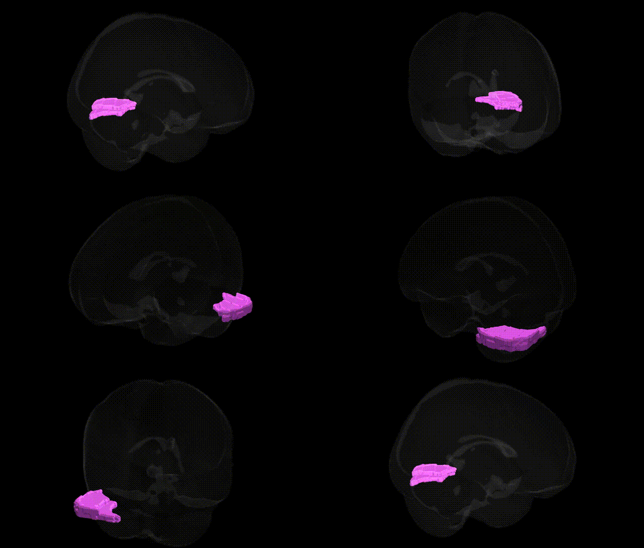
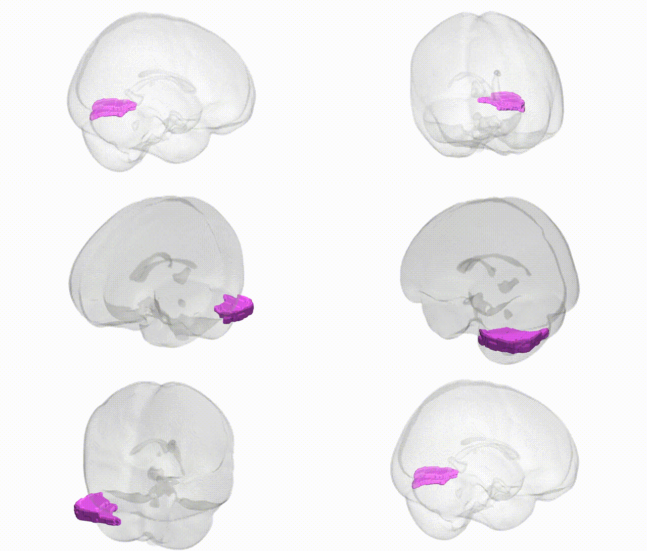
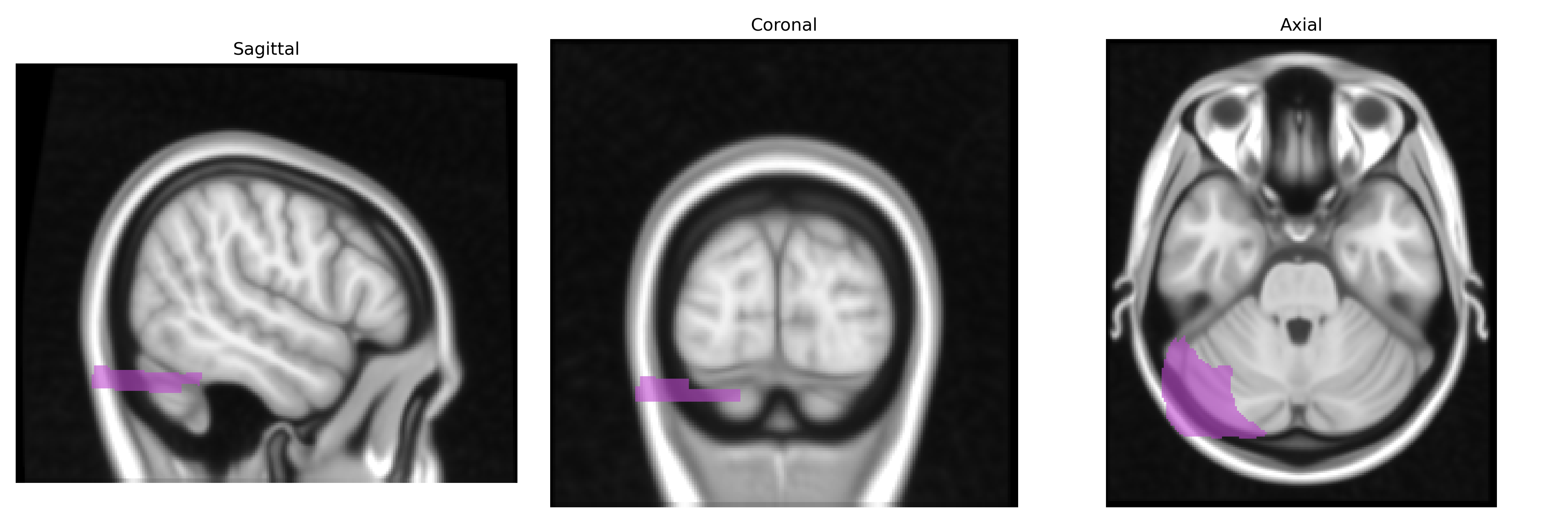
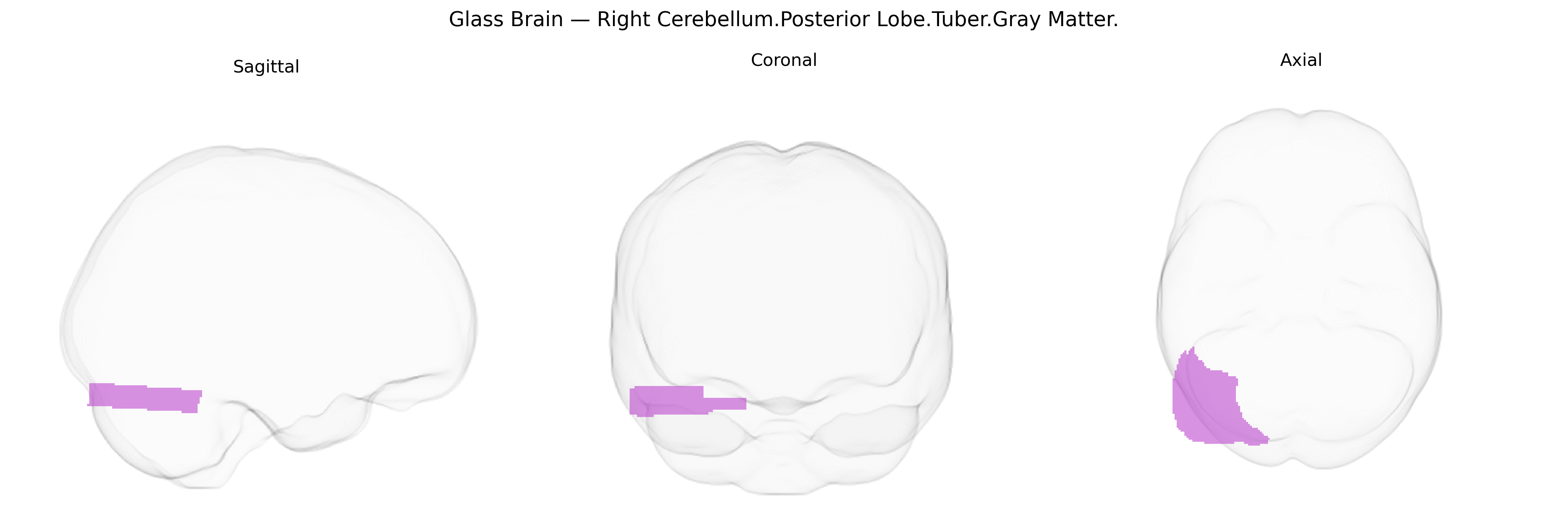
The bilateral Right Cerebellum Posterior Lobe Tuber (gray matter) corresponds to a portion of lobule VII (often within Crus II) on the inferior surface of the cerebellar hemisphere, forming part of the posterior lobe that participates in higher-order motor coordination and cognitive processing. As gray matter, it contains densely packed neuronal cell bodies within the cerebellar cortex, organized into the characteristic three-layered structure (molecular, Purkinje cell, and granular layers) that modulates output to the deep cerebellar nuclei. Functionally, this region is implicated in fine-tuning voluntary movements, motor learning, and timing, and has been associated via functional imaging with aspects of executive function, language, and visuospatial processing through its connections with cerebral association cortices and thalamic relay nuclei. There is no direct link for this specific subregion; a related structure is the Cerebellum.
Genetic associations specifically targeting the bilateral right cerebellum posterior lobe tuber gray matter, as defined in the Talairach 1 mm atlas, are sparse, but broader imaging-genetics and GWAS literature implicates overlapping cerebellar posterior lobe territories in several traits and disorders. Large-scale brain MRI GWAS (e.g., ENIGMA and UK Biobank–based studies) have identified common variants in genes related to neurodevelopment (such as KIAA0586, RELN, and FOXP2 pathways), axon guidance, and synaptic organization that influence cerebellar gray-matter volume and surface-based morphometry, including in posterior lobules. Polygenic risk scores for schizophrenia, bipolar disorder, major depression, and autism spectrum disorder show associations with altered cerebellar morphology and connectivity, with posterior lobe involvement in cognitive and affective circuits. Variants in genes involved in synaptic plasticity and glutamatergic signaling (e.g., GRM and CACNA family members) have been linked to cerebellar structural and functional differences in disorders such as schizophrenia, ataxias, and developmental coordination disorder, and several spinocerebellar ataxia genes (e.g., ATXN1, ATXN2, CACNA1A) cause degenerative changes that prominently involve posterior cerebellar regions, although not confined to the tuber. Additionally, GWAS of cognitive performance and educational attainment highlight loci associated with cerebellar volume and functional connectivity, reinforcing the role of posterior cerebellar territories in higher cognition and suggesting that multiple small-effect common variants collectively modulate gray-matter properties in this region, rather than any single locus being uniquely tied to the tuber.
Overview generated by GPT-4o (2026).
Region ID: 81
Hemisphere: bilateral
Atlas: Talairach labels 1mm
Full Quality Version: Download MP4
Full Quality Version: Download MP4
Wali Sidiqyar*, Gaurav Rudravaram*, Elyssa M. McMaster, Trent M. Schwartz, Adam M. Saunders, Kurt G. Schilling, Bennett A. Landman "Introducing SPINS: A Shared Public Visualization Library of Neuroanatomical Structures." Medical Imaging with Deep Learning- short paper
This resource is licensed under CC0 1.0 Universal (Public Domain).