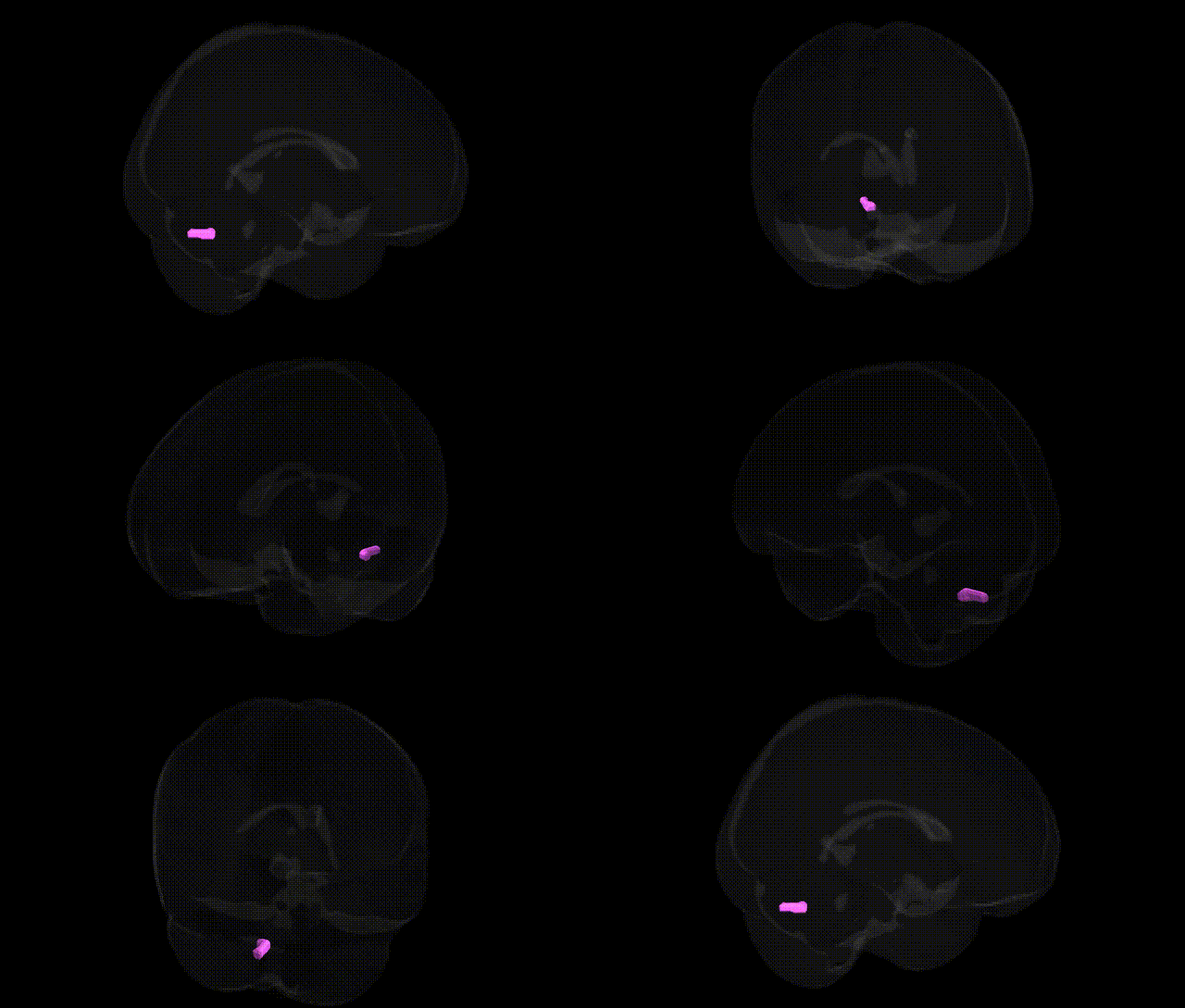
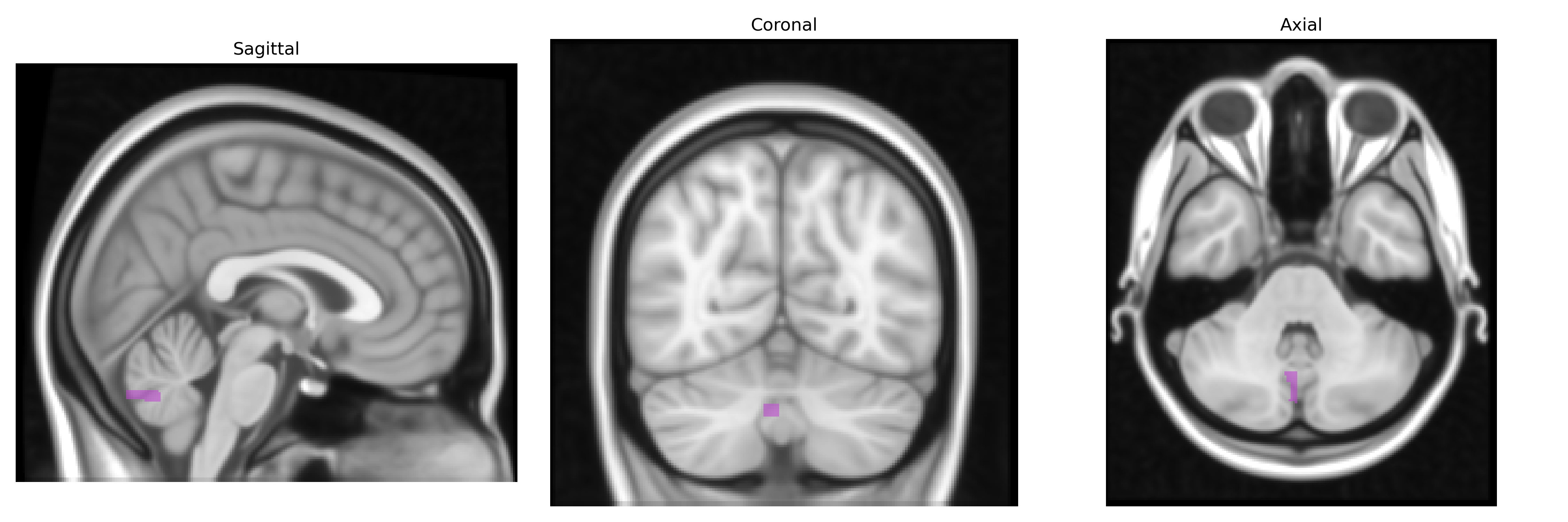
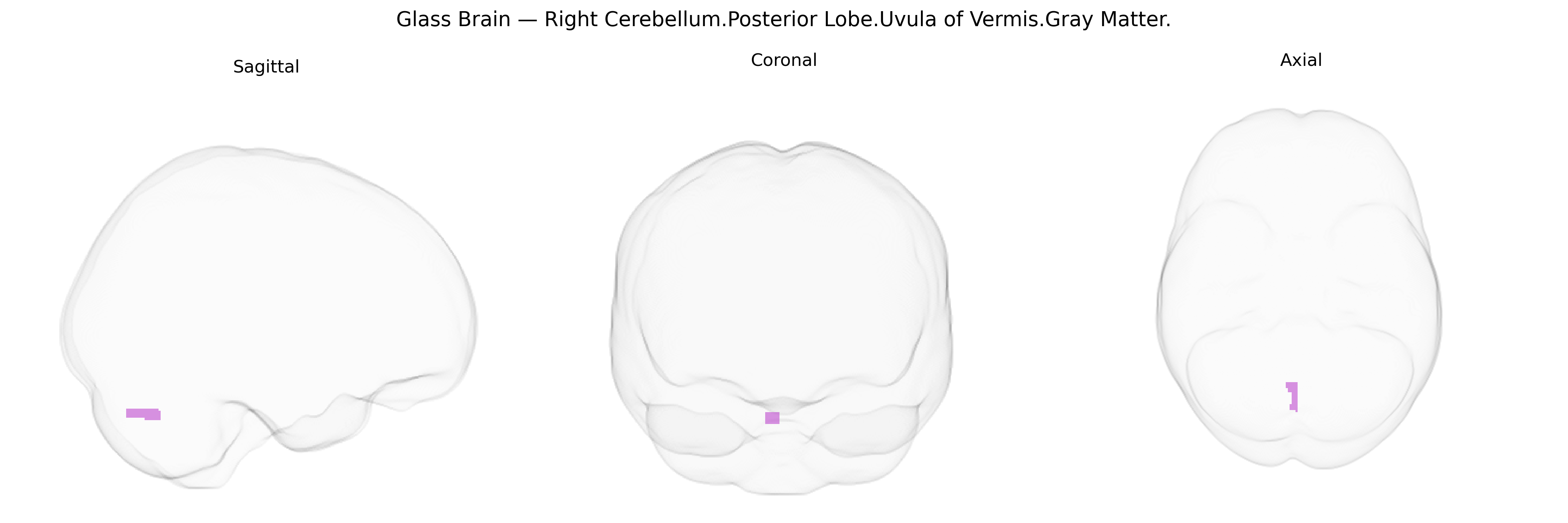
The bilateral Right Cerebellum, Posterior Lobe, Uvula of Vermis, Gray Matter corresponds to a midline vermal structure within the posterior lobe of the cerebellum, composed predominantly of cortical gray matter containing densely packed neurons that contribute to cerebellar microcircuitry. The uvula of the vermis participates in the regulation of balance, posture, and coordination of axial and proximal limb movements, receiving afferent input from spinal, vestibular, and cortical sources and projecting to deep cerebellar nuclei that influence brainstem and thalamocortical motor pathways. It is part of the functional domain involved in fine-tuning motor execution and adaptive motor learning, and may also contribute to oculomotor and vestibular control. There is no direct Wikipedia article for this specific subregion; see the related structure Cerebellar vermis.
The bilateral right cerebellum posterior lobe, specifically the uvula of the vermis gray matter (Talairach 1 mm Atlas), is part of the midline cerebellar vermis implicated in motor coordination, affect regulation, and cognitive processing, and genetic associations involving this region typically arise from imaging genetics and cerebellar volume GWAS rather than label-specific studies. Large-scale GWAS of cerebellar volume and morphology (e.g., studies identifying variants in genes such as LRP1B, PAPPA, KATNAL2, and loci on chromosomes 1, 3, 6, and 16) have shown that common variants influence regional cerebellar structure, with vermis and posterior lobe measures contributing to polygenic architecture; however, these findings usually report broader lobular or vermal measures rather than the uvula subregion. The cerebellar vermis, including posterior portions overlapping the uvula, has been repeatedly implicated in neurodevelopmental and psychiatric conditions—such as autism spectrum disorder, schizophrenia, bipolar disorder, and major depression—where case-control and family-based MRI studies link disorder-associated risk genes (e.g., CACNA1C, DISC1, CNTNAP2, SHANK3, and synaptic genes identified in ASD and schizophrenia GWAS) to altered vermis volume or functional connectivity, though not uniquely to the uvula. In ADHD, OCD, and mood disorders, polygenic risk scores and GWAS of symptom dimensions have been associated with structural and functional variation in the posterior cerebellum and vermis in imaging-genetic studies, suggesting that many brain-relevant loci (e.g., in glutamatergic, GABAergic, and neurodevelopmental pathways) exert pleiotropic effects that include this region. Additionally, rare variant and copy number variant studies in syndromic cerebellar malformations and developmental disorders (e.g., Joubert-spectrum genes like AHI1, CEP290, and other ciliary and axonal guidance genes) demonstrate that mutations affecting cerebellar midline development can involve the vermis and adjacent uvular territory, though these are typically described at the level of vermis hypoplasia rather than discrete Talairach-defined parcels. Overall, genetic findings tied to this specific atlas-defined uvula region are indirect and inferred from broader vermis/posterior lobe cerebellar phenotypes, with no current GWAS reporting loci uniquely for the “Right Cerebellum.Posterior Lobe.Uvula of Vermis.Gray Matter” label, but multiple neuropsychiatric and neurodevelopmental risk genes appear to influence its structure and function as part of the cerebellar vermis network.
Overview generated by GPT-4o (2026).
Region ID: 68
Hemisphere: bilateral
Atlas: Talairach labels 1mm
Full Quality Version: Download MP4
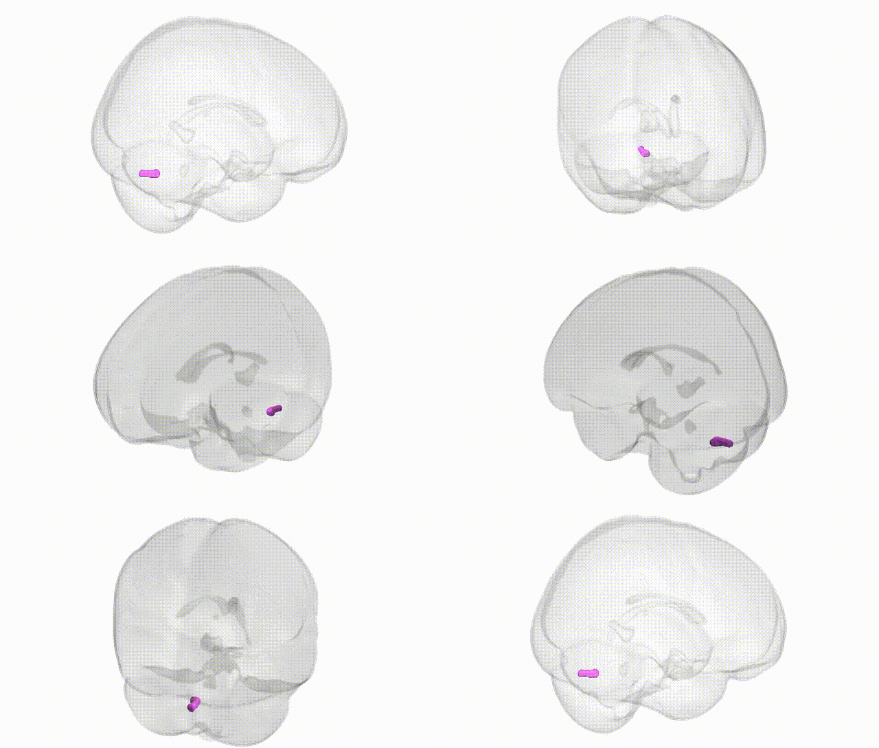
Full Quality Version: Download MP4
Wali Sidiqyar*, Gaurav Rudravaram*, Elyssa M. McMaster, Trent M. Schwartz, Adam M. Saunders, Kurt G. Schilling, Bennett A. Landman "Introducing SPINS: A Shared Public Visualization Library of Neuroanatomical Structures." Medical Imaging with Deep Learning- short paper
This resource is licensed under CC0 1.0 Universal (Public Domain).