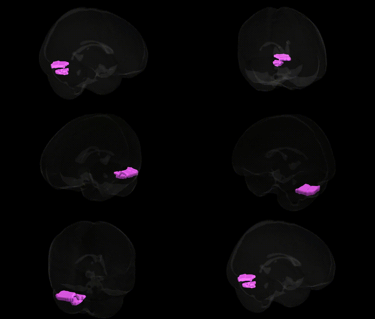
The bilateral Right Cerebellum Posterior Lobe Uvula (gray matter) is a vermal region of the posterior lobe of the cerebellum, located in the medial portion of the inferior cerebellar vermis. It contributes to the fine-tuning of motor coordination, balance, and posture by integrating vestibular, proprioceptive, and somatosensory inputs, and is involved in the regulation of axial musculature and eye movements. Functionally, this gray matter region participates in cerebellar circuitry that modulates motor output via connections with deep cerebellar nuclei and brainstem motor centers, and may also play a role in aspects of cognitive and affective processing attributed to the cerebellar vermis. There is no direct link for the uvula of the cerebellum; a related structure is the Cerebellum.
The bilateral Right Cerebellum.Posterior Lobe.Uvula.Gray Matter, as defined in the Talairach 1 mm atlas, falls within the posterior cerebellar vermis and adjacent hemispheric cortex, a region generally implicated in motor coordination, affective regulation, and aspects of cognition; however, most large-scale genetic imaging studies and GWAS have not isolated the uvula subregion specifically, instead reporting broader associations for the posterior cerebellum or total cerebellar gray matter. Genome-wide association studies of brain structure (e.g., ENIGMA and UK Biobank–based analyses) have identified common variants in genes involved in neurodevelopment, synaptic function, and axonal guidance (such as those near genes like KIAA0586, RELN, and DCC) that influence overall cerebellar volume or lobule-level anatomy, but these findings typically map to larger lobular parcels encompassing lobules VIII–X rather than the uvula alone. Genetic studies of neurodevelopmental and psychiatric disorders—including autism spectrum disorder, schizophrenia, bipolar disorder, and major depression—have repeatedly implicated cerebellar gray matter alterations, especially in posterior lobules, and polygenic risk scores for these conditions often correlate with differences in cerebellar morphology or connectivity, yet the spatial resolution of most imaging-genetic datasets remains insufficient to assign robust, uvula-specific genetic risk. Similarly, GWAS of motor skills, educational attainment, and cognitive performance show associations with cerebellar metrics at a lobular or global level, suggesting shared polygenic influences on cortico-cerebellar networks without pinpointing distinct loci uniquely tied to the right posterior-lobe uvula. Overall, current evidence supports a polygenic architecture affecting the posterior cerebellum as part of distributed motor-cognitive circuits, but no well-replicated GWAS signals or single-gene disorders can be confidently localized to the bilateral Right Cerebellum.Posterior Lobe.Uvula.Gray Matter region at the fine anatomical resolution implied by the Talairach atlas.
Overview generated by GPT-4o (2026).
Region ID: 64
Hemisphere: bilateral
Atlas: Talairach labels 1mm
Full Quality Version: Download MP4
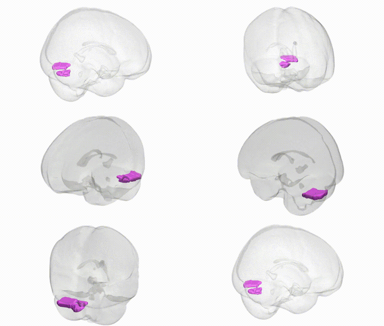
Full Quality Version: Download MP4
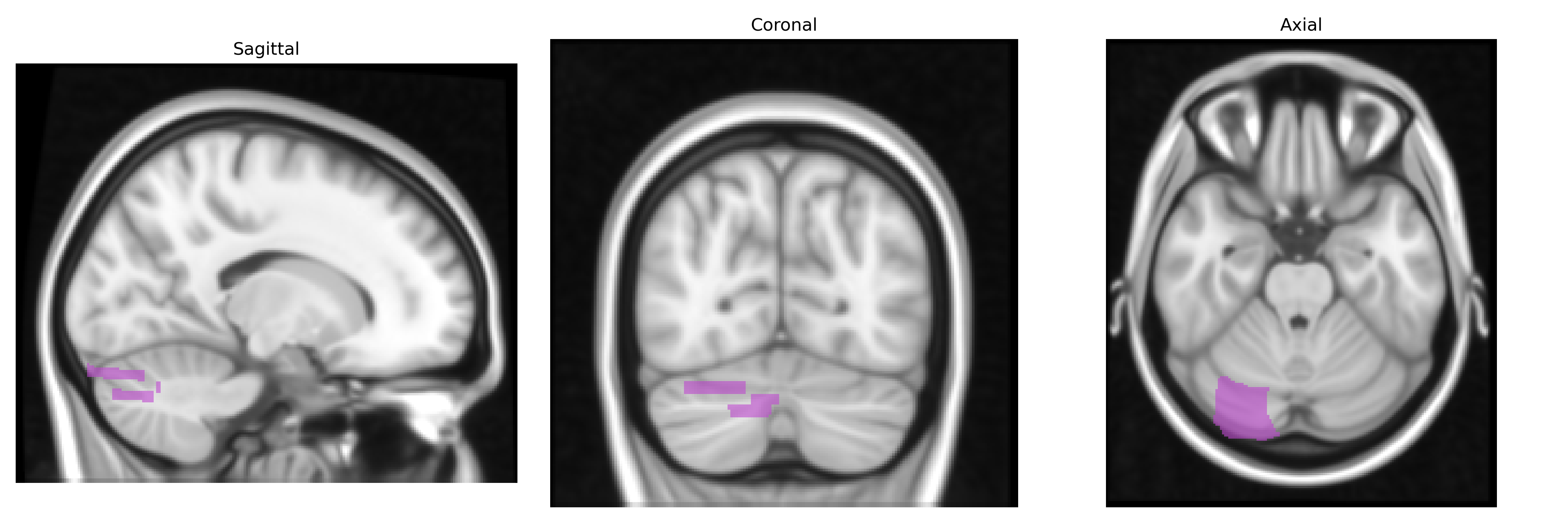
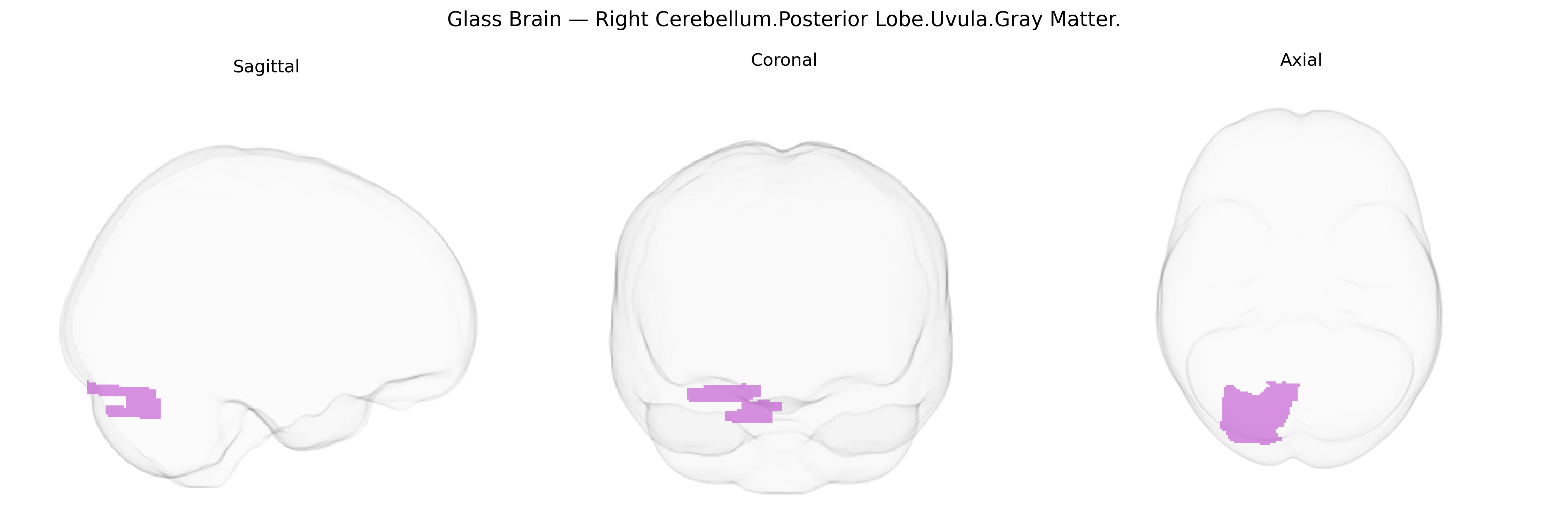
Wali Sidiqyar*, Gaurav Rudravaram*, Elyssa M. McMaster, Trent M. Schwartz, Adam M. Saunders, Kurt G. Schilling, Bennett A. Landman "Introducing SPINS: A Shared Public Visualization Library of Neuroanatomical Structures." Medical Imaging with Deep Learning- short paper
This resource is licensed under CC0 1.0 Universal (Public Domain).