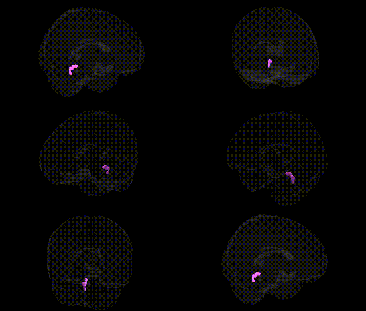
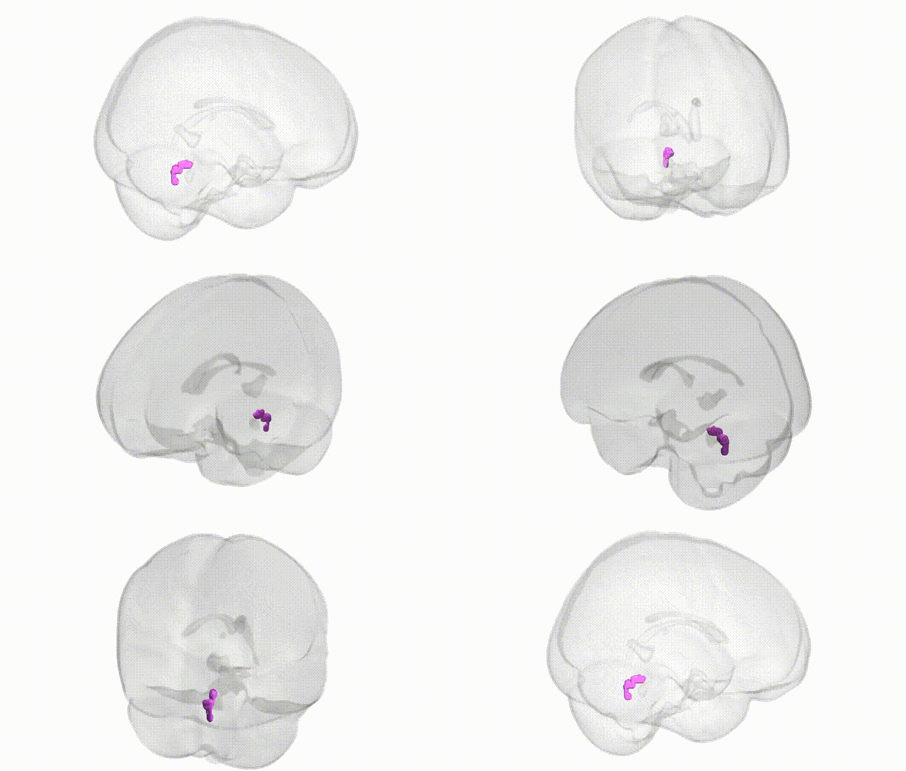
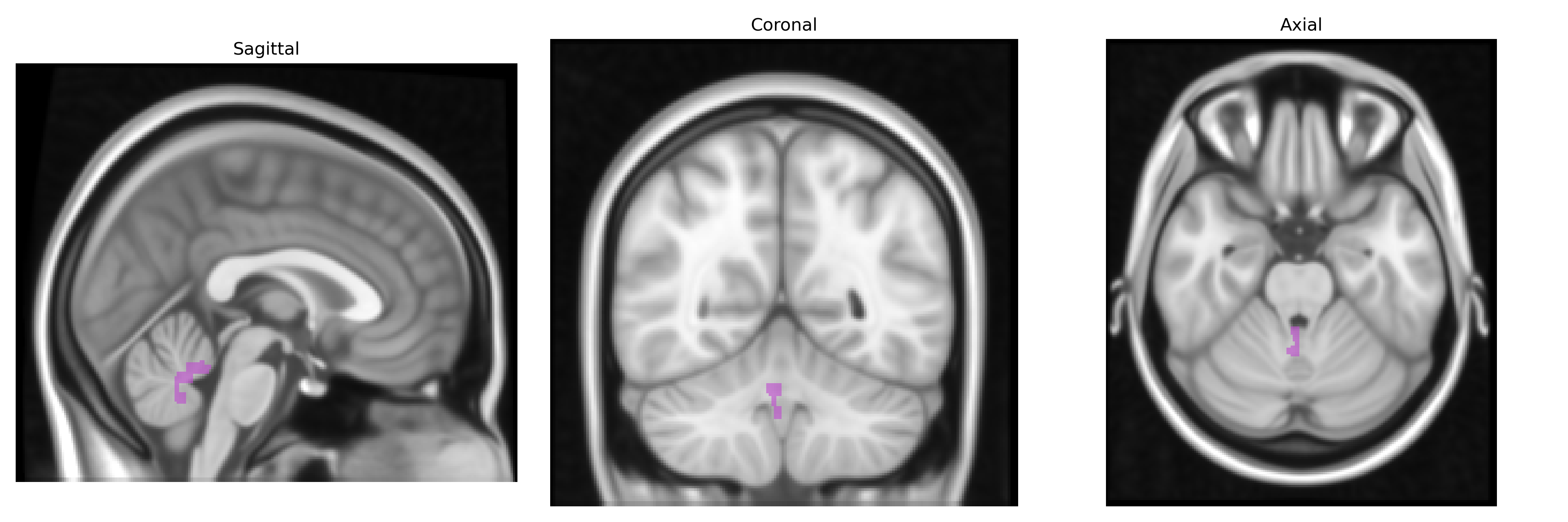
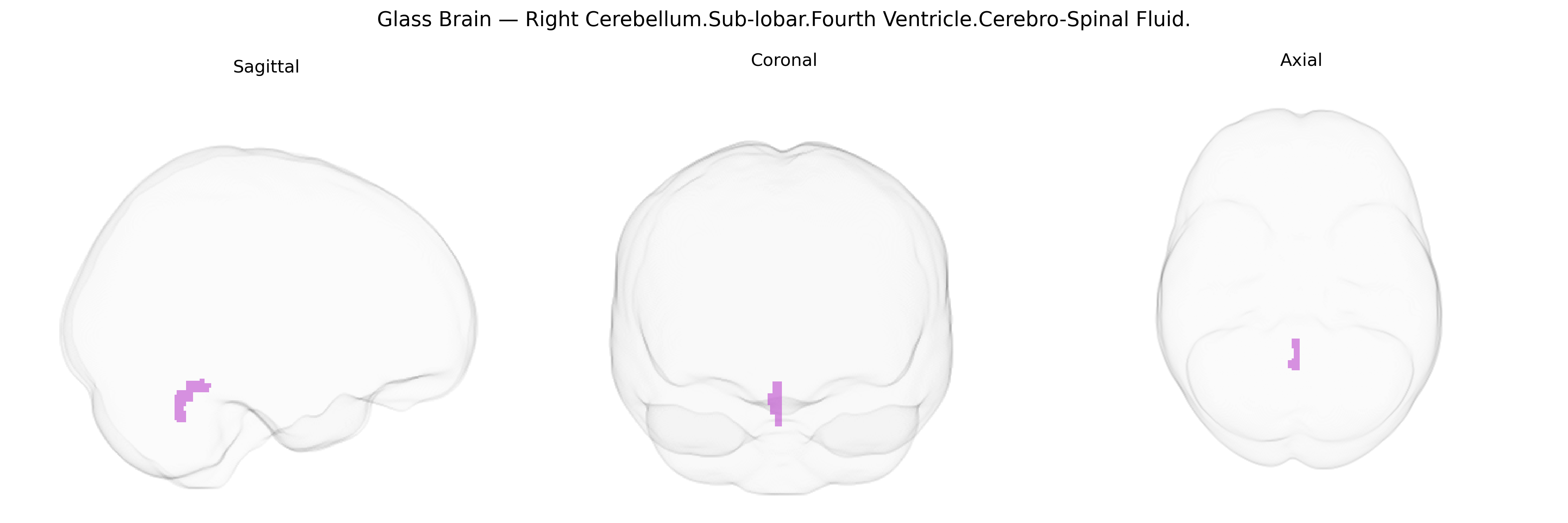
The bilateral Right Cerebellum.Sub-lobar.Fourth Ventricle.Cerebro-Spinal Fluid region in the Talairach 1 mm Atlas denotes voxels located within the cerebrospinal fluid (CSF)–filled cavity of the fourth ventricle, anatomically bordered ventrally by the pons and dorsally by the cerebellum, particularly its right hemispheric structures. This space forms part of the ventricular system that facilitates circulation of CSF from the cerebral aqueduct to the central canal of the spinal cord and into the subarachnoid space via the median and lateral apertures, contributing to mechanical protection, metabolic waste clearance, and maintenance of intracranial pressure and homeostasis for adjacent brainstem and cerebellar tissue. There is no direct link for this compound label; a related structure entry is: Fourth ventricle.
Genetic association studies rarely target the bilateral “Right Cerebellum.Sub-lobar.Fourth Ventricle.Cerebro-Spinal Fluid” label from the Talairach 1 mm atlas as a discrete region, but related work on cerebellar volume, posterior fossa structures, and ventricular/CSF measures provides indirect evidence. GWAS of global and regional brain volumes (e.g., ENIGMA, UK Biobank) have identified loci in genes such as KCNK2, PAPPA, and VCAN associated with total cerebellar volume and posterior fossa morphology, while large-scale analyses of ventricular size and intracranial CSF volume implicate genes involved in neurodevelopment, axon guidance, and extracellular matrix (e.g., genes near APOE, WWOX, and MAPT in some cohorts). Variants influencing cerebellar and ventricular traits often overlap with risk loci for neurodevelopmental and neuropsychiatric disorders, including schizophrenia, bipolar disorder, autism spectrum disorder, and major depression, where altered cerebellar structure and fourth-ventricle/CSF morphology have been reported. Additionally, genes associated with hydrocephalus and posterior fossa malformations (such as L1CAM and certain ciliopathy genes) can alter fourth-ventricle size and CSF dynamics, although these findings typically relate to broader ventricular and cerebellar anomalies rather than the specific Talairach-defined sub-lobar CSF region. Overall, current genetic evidence links variants affecting cerebellar development, ventricular size, and CSF regulation to structural differences in regions encompassing the fourth ventricle, but direct GWAS focused specifically on the Talairach “Right Cerebellum.Sub-lobar.Fourth Ventricle.CSF” label are lacking.
Overview generated by GPT-4o (2026).
Region ID: 69
Hemisphere: bilateral
Atlas: Talairach labels 1mm
Full Quality Version: Download MP4
Full Quality Version: Download MP4
Wali Sidiqyar*, Gaurav Rudravaram*, Elyssa M. McMaster, Trent M. Schwartz, Adam M. Saunders, Kurt G. Schilling, Bennett A. Landman "Introducing SPINS: A Shared Public Visualization Library of Neuroanatomical Structures." Medical Imaging with Deep Learning- short paper
This resource is licensed under CC0 1.0 Universal (Public Domain).