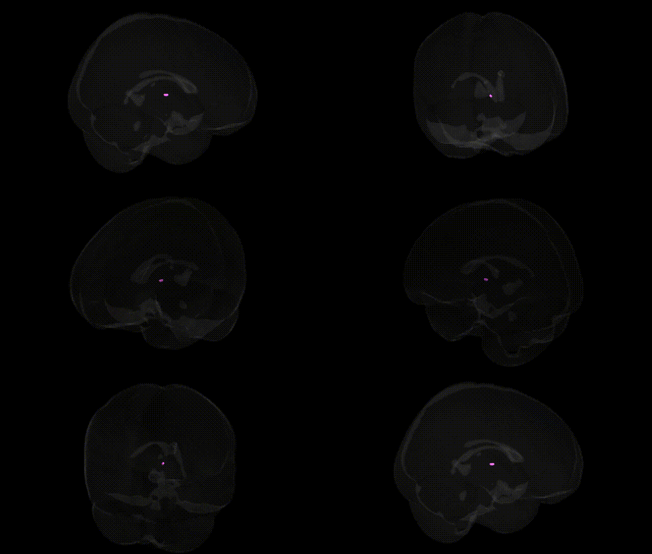
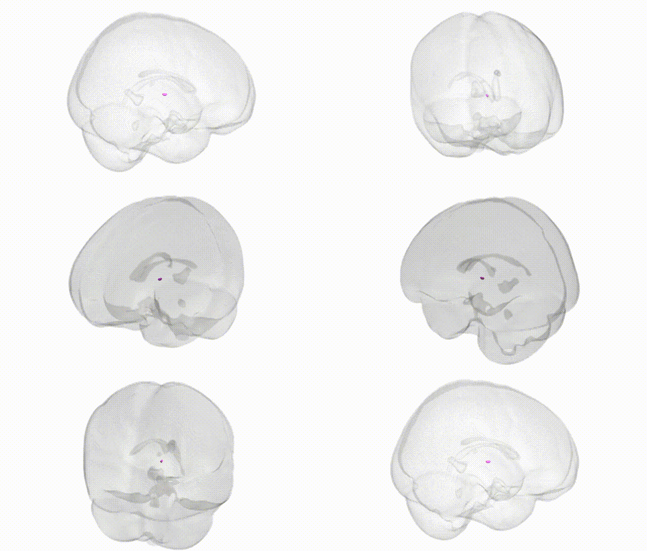
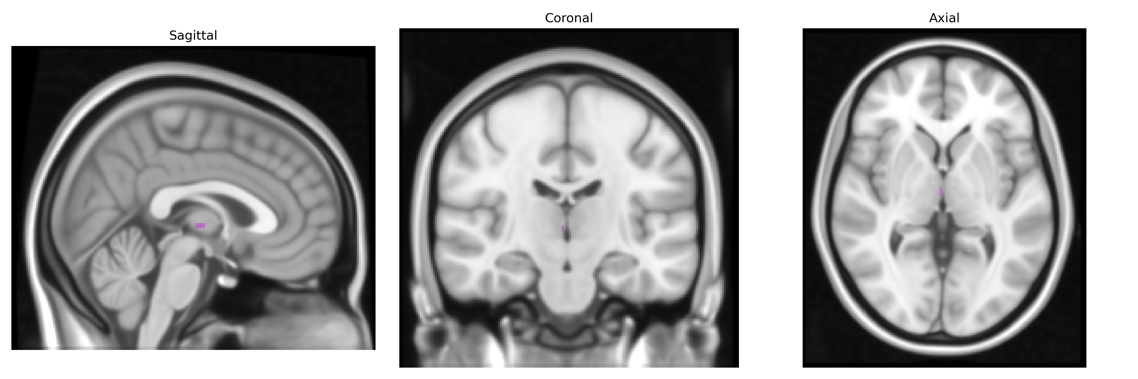
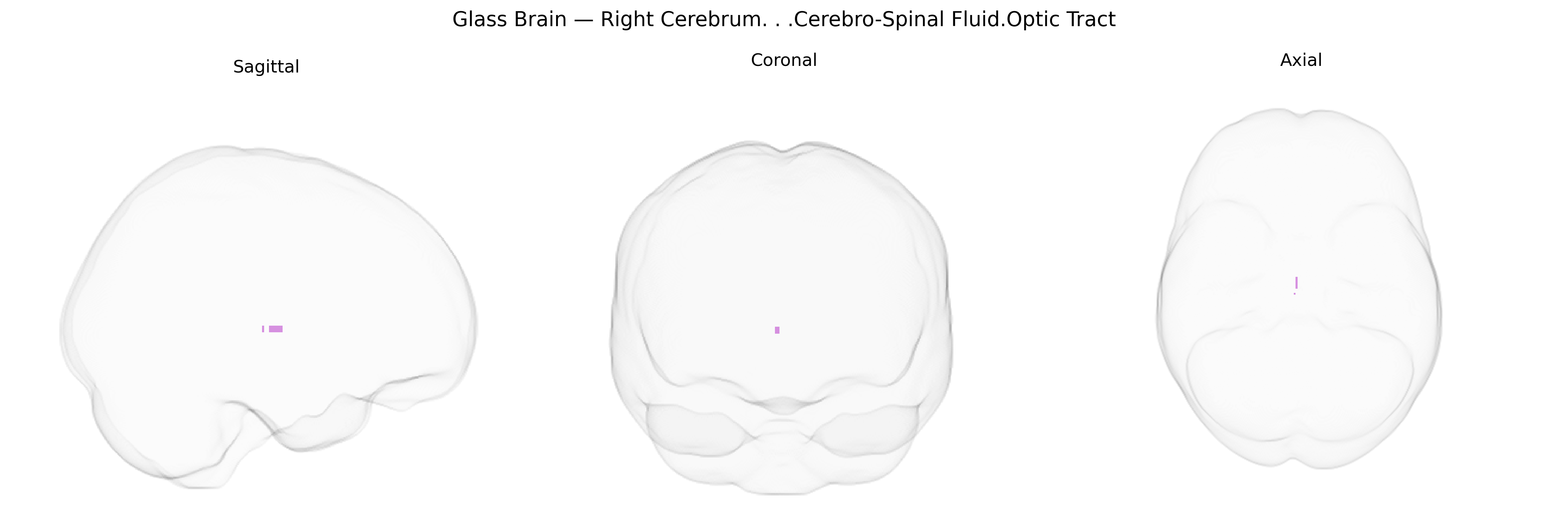
The bilateral Right Cerebrum / Cerebro-Spinal Fluid / Optic Tract region, as labeled in the Talairach 1 mm atlas, encompasses cerebrospinal fluid spaces adjacent to the right cerebral hemisphere and includes the vicinity of the optic tract, a major white matter pathway of the visual system. The optic tract carries visual information from the optic chiasm to the lateral geniculate nucleus, superior colliculus, and other midbrain and thalamic targets, conveying signals that are critical for conscious visual perception, reflexive eye movements, and visual attention. Located along the ventrolateral surface of the diencephalon, the optic tract runs posteriorly and slightly laterally from the chiasm, tightly associated with surrounding CSF-containing cisterns and the basal surface of the right cerebrum. There is no direct link for this composite label; a related structure is the Optic tract.
The optic tract region within the right cerebrum, adjacent to cerebrospinal fluid spaces, has been implicated in numerous imaging-genetics and GWAS studies focused on visual system integrity, white-matter microstructure, and neuro-ophthalmologic disease. Variants in genes critical for axonal guidance and myelination (such as ROBO1, PLP1, LINGO1, and MAG) have been associated with diffusion tensor imaging measures and structural variation in optic pathways, including the optic tract, while loci influencing intracranial volume and global white-matter properties (e.g., near HMGA2 and IGF1) also contribute to interindividual differences in this region’s volume and connectivity. GWAS of glaucoma and optic neuropathies have identified risk variants in genes such as MYOC, OPTN, WDR36, and SIX6 that affect retinal ganglion cell survival and downstream optic tract integrity, with corresponding changes detectable in MRI-based measures of optic tract structure. In neurodegenerative and demyelinating disorders, including multiple sclerosis, Leber hereditary optic neuropathy (MT-ND genes), and autosomal dominant optic atrophy (OPA1), pathogenic variants lead to degeneration of optic tract fibers, and imaging studies consistently show atrophy or altered microstructure in this Talairach-defined region. Large consortia such as ENIGMA and UK Biobank have further demonstrated that polygenic scores related to neurodevelopment, cognition, and psychiatric risk (schizophrenia, major depression, and bipolar disorder) correlate with white-matter integrity in visual pathways, including the optic tract, highlighting this region as a genetically influenced node linking visual processing, neurodegeneration, and broader brain-wide connectivity.
Overview generated by GPT-4o (2026).
Region ID: 639
Hemisphere: bilateral
Atlas: Talairach labels 1mm
Full Quality Version: Download MP4
Full Quality Version: Download MP4
Wali Sidiqyar*, Gaurav Rudravaram*, Elyssa M. McMaster, Trent M. Schwartz, Adam M. Saunders, Kurt G. Schilling, Bennett A. Landman "Introducing SPINS: A Shared Public Visualization Library of Neuroanatomical Structures." Medical Imaging with Deep Learning- short paper
This resource is licensed under CC0 1.0 Universal (Public Domain).