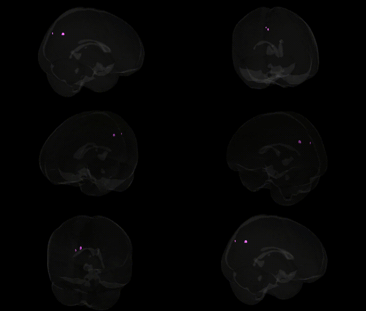
The bilateral Right Cerebrum, Cuneus, corresponds to the cuneus region of the occipital lobe, a wedge-shaped cortical area located on the medial surface of the hemisphere between the parieto-occipital sulcus and the calcarine sulcus. It is primarily involved in early-stage visual processing, including basic features such as orientation, spatial frequency, and some aspects of visual attention, and contributes to the integration of visual information from the primary visual cortex (V1) with higher-order visual areas. The cuneus participates in processing input from the contralateral visual field and supports functions related to visual perception, visuospatial orientation, and, in some networks, aspects of visual imagery and attention. Cuneus
The bilateral right cuneus, part of the occipital cortex implicated in primary and secondary visual processing, has been repeatedly implicated in imaging–genetics and GWAS-derived endophenotype studies, where variation in cortical thickness, surface area, or activation has been associated with common variants in genes involved in neurodevelopment, synaptic signaling, and myelination (for example, large ENIGMA and UK Biobank analyses highlight loci near or within HMGA2, RSPO3, TBR1, and microtubule- or axon-guidance related genes for occipital and visual cortex morphology and connectivity). Polygenic risk for schizophrenia, bipolar disorder, and major depressive disorder shows associations with structural and functional alterations in the cuneus, and specific risk loci (e.g., in CACNA1C, MIR137-regulated networks, and complement pathway genes) have been linked to occipital/cuneus volume or activation patterns in case–control imaging–genetics cohorts. GWAS of cognitive and visual traits, including general cognitive ability, visual attention, and reading-related measures, have reported cuneus involvement as a mediating region for variants in neurodevelopmental transcription factors and axonal development genes, while studies of neurodegenerative disorders such as Alzheimer’s disease and multiple sclerosis identify cuneus atrophy or lesion load as imaging phenotypes partially explained by APOE and immune-related risk loci, suggesting that genetic influences on this region contribute to vulnerability in both psychiatric and neurodegenerative phenotypes.
Overview generated by GPT-4o (2026).
Region ID: 951
Hemisphere: bilateral
Atlas: Talairach labels 1mm
Full Quality Version: Download MP4
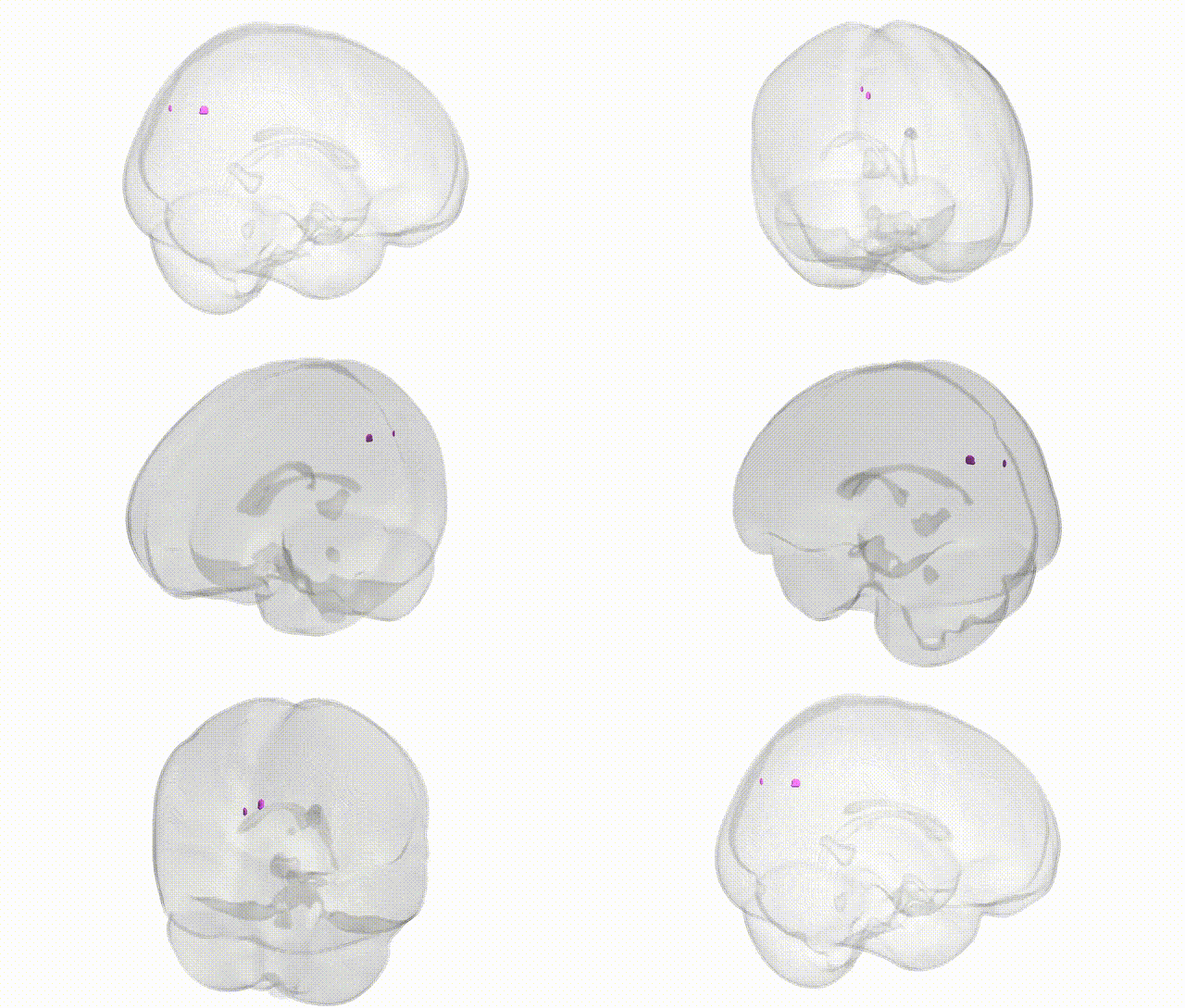
Full Quality Version: Download MP4
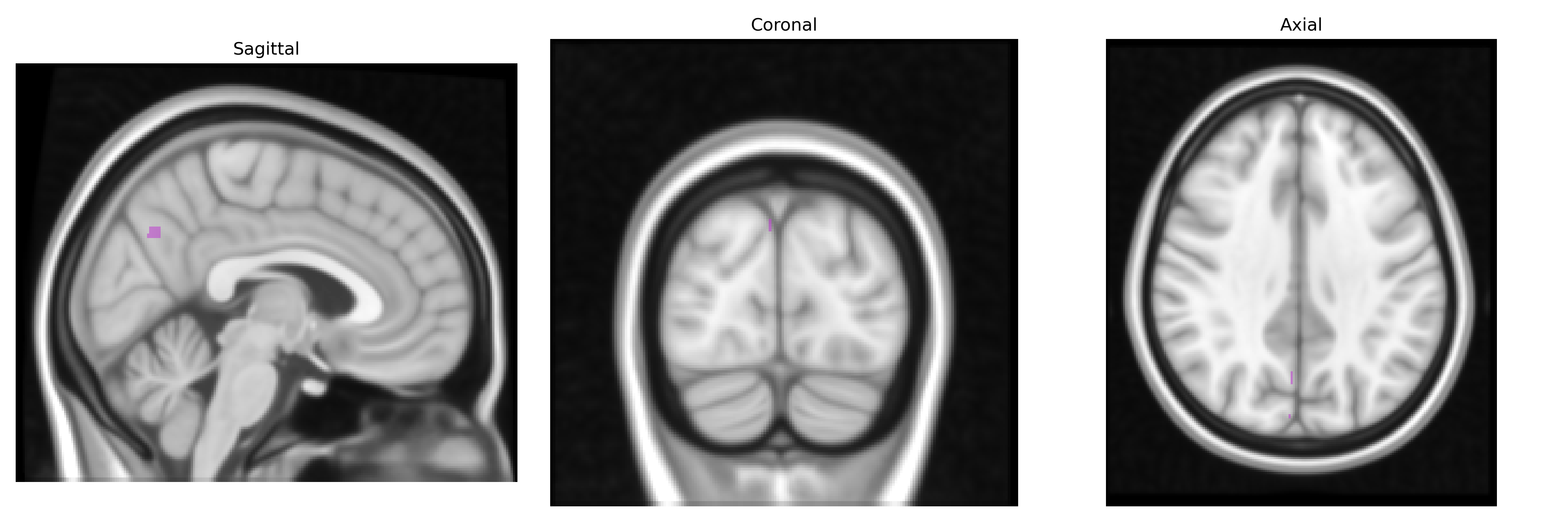
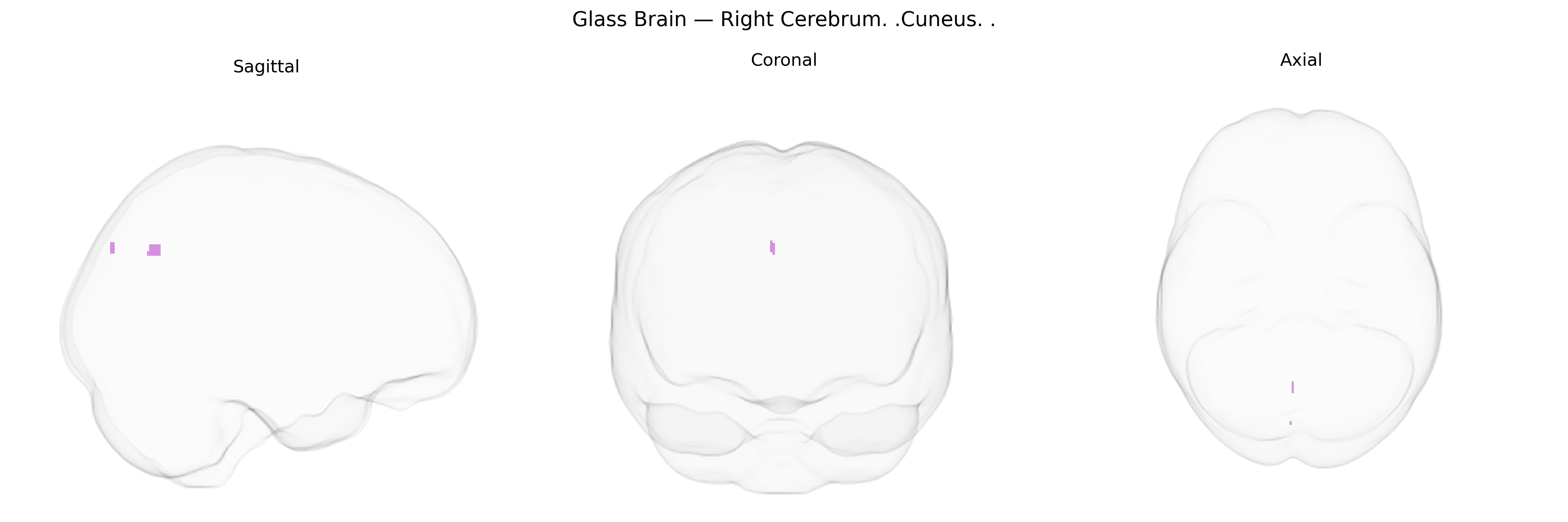
Wali Sidiqyar*, Gaurav Rudravaram*, Elyssa M. McMaster, Trent M. Schwartz, Adam M. Saunders, Kurt G. Schilling, Bennett A. Landman "Introducing SPINS: A Shared Public Visualization Library of Neuroanatomical Structures." Medical Imaging with Deep Learning- short paper
This resource is licensed under CC0 1.0 Universal (Public Domain).